 Movie
Movie Controller

[English] 日本語
 Yorodumi
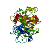
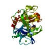
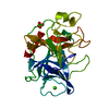
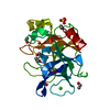
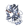
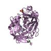
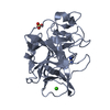
- PDB-1est: THE ATOMIC STRUCTURE OF CRYSTALLINE PORCINE PANCREATIC ELASTASE A... -
+ Open data

- Basic information
| Entry | Database: PDB / ID: 1est | ||||||
|---|---|---|---|---|---|---|---|
| Title | THE ATOMIC STRUCTURE OF CRYSTALLINE PORCINE PANCREATIC ELASTASE AT 2.5 ANGSTROMS RESOLUTION. COMPARISONS WITH THE STRUCTURE OF ALPHA-CHYMOTRYPSIN | ||||||
 Components Components | PORCINE PANCREATIC ELASTASE | ||||||
 Keywords Keywords | HYDROLASE / HYDROLASE (SERINE PROTEINASE) | ||||||
| Function / homology |  Function and homology information Function and homology informationpancreatic elastase / serine-type endopeptidase activity / proteolysis / : / metal ion binding Similarity search - Function | ||||||
| Biological species |  | ||||||
| Method |  X-RAY DIFFRACTION / Resolution: 2.5 Å X-RAY DIFFRACTION / Resolution: 2.5 Å | ||||||
 Authors Authors | Sawyer, L. / Shotton, D.M. / Watson, H.C. | ||||||
 Citation Citation | Journal: J.Mol.Biol. / Year: 1978 Title: The atomic structure of crystalline porcine pancreatic elastase at 2.5 A resolution: comparisons with the structure of alpha-chymotrypsin. Authors: Sawyer, L. / Shotton, D.M. / Campbell, J.W. / Wendell, P.L. / Muirhead, H. / Watson, H.C. / Diamond, R. / Ladner, R.C. #1: Journal: Biochem.Biophys.Res.Commun. / Year: 1973Title: Atomic Coordinates for Tosyl-Elastase Authors: Sawyer, L. / Shotton, D.M. / Watson, H.C. #2: Journal: Nature / Year: 1970Title: Amino-Acid Sequence of Porcine Pancreatic Elastase and its Homologies with Other Serine Proteinases Authors: Shotton, D.M. / Hartley, B.S. #3: Journal: Nature / Year: 1970Title: Three-Dimensional Fourier Synthesis of Tosyl-Elastase at 3.5 Angstroms Resolution Authors: Watson, H.C. / Shotton, D.M. / Cox, J.M. / Muirhead, H. #4: Journal: Nature / Year: 1970Title: Three Dimensional Structure of Tosyl-Elastase Authors: Shotton, D.M. / Watson, H.C. | ||||||
| History |
| ||||||
| Remark 700 | SHEET THE TWO SEVEN STRANDED SHEETS IN THIS STRUCTURE ARE REALLY SIX STRANDED BETA BARRELS - THIS ...SHEET THE TWO SEVEN STRANDED SHEETS IN THIS STRUCTURE ARE REALLY SIX STRANDED BETA BARRELS - THIS IS DENOTED BY THE FIRST STRAND RECURRING AS THE LAST STRAND. |
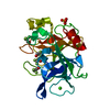
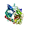
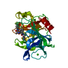
- Structure visualization
| Structure viewer | Molecule: MolmilJmol/JSmol |
|---|
- Downloads & links
-Download
| PDBx/mmCIF format | 1est.cif.gz | 54.2 KB | Display | PDBx/mmCIF format |
|---|---|---|---|---|
| PDB format | pdb1est.ent.gz | 37.9 KB | Display | PDB format |
| PDBx/mmJSON format | 1est.json.gz | Tree view | PDBx/mmJSON format | |
| Others |  Other downloads Other downloads |
-Validation report
| Arichive directory | https://data.pdbj.org/pub/pdb/validation_reports/es/1estftp://data.pdbj.org/pub/pdb/validation_reports/es/1est | HTTPS FTP |
|---|
-Related structure data
| Similar structure data |
|---|
-Links
 PDBj
PDBj

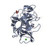
- Assembly
| Deposited unit | 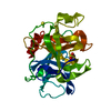
| ||||||||
|---|---|---|---|---|---|---|---|---|---|
| 1 |
| ||||||||
| Unit cell |
| ||||||||
| Atom site foot note | 1: RESIDUE SER 195 (THE CATALYTICALLY ACTIVE SERINE ) HAS BEEN TOSYLATED IN THIS STRUCTURE. COORDINATES FOR THE TOSYL GROUP ARE GIVEN IN THE HETATM RECORDS BELOW. |
-Components
| #1: Protein | Mass: 25928.031 Da / Num. of mol.: 1 Source method: isolated from a genetically manipulated source Source: (gene. exp.) |
|---|---|
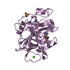
| #2: Chemical | ChemComp-SO4 /   Mass: 96.063 Da / Num. of mol.: 1 / Source method: obtained synthetically / Formula: SO4 Mass: 96.063 Da / Num. of mol.: 1 / Source method: obtained synthetically / Formula: SO4 |
| #3: Chemical | ChemComp-TSU / 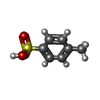  Mass: 172.202 Da / Num. of mol.: 1 / Source method: obtained synthetically / Formula: C7H8O3S Mass: 172.202 Da / Num. of mol.: 1 / Source method: obtained synthetically / Formula: C7H8O3S |
| #4: Water | ChemComp-HOH /  Mass: 18.015 Da / Num. of mol.: 25 / Source method: isolated from a natural source / Formula: H2O Mass: 18.015 Da / Num. of mol.: 25 / Source method: isolated from a natural source / Formula: H2O |
| Has protein modification | Y |
-Experimental details
-Experiment
| Experiment | Method: X-RAY DIFFRACTION |
|---|
- Sample preparation
| Crystal | Density Matthews: 2.17 Å3/Da / Density % sol: 43.42 % | ||||||||||||||||||||
|---|---|---|---|---|---|---|---|---|---|---|---|---|---|---|---|---|---|---|---|---|---|
| Crystal grow | *PLUS Temperature: 2 ℃ / pH: 5 / Method: unknown | ||||||||||||||||||||
| Components of the solutions | *PLUS
|
-Data collection
| Radiation | Scattering type: x-ray |
|---|---|
| Radiation wavelength | Relative weight: 1 |
- Processing
| Refinement | Highest resolution: 2.5 Å Details: COORDINATES FOR THE 27 WATER MOLECULES AND 1 SULPHATE ION WERE TAKEN FROM THE ARTICLE CITED AS REFERENCE 1. | ||||||||||||
|---|---|---|---|---|---|---|---|---|---|---|---|---|---|
| Refinement step | Cycle: LAST / Highest resolution: 2.5 Å
|
