 Movie
Movie Controller

[English] 日本語
 Yorodumi
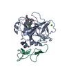
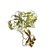
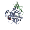
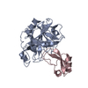
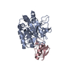
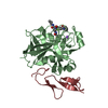
- PDB-1fle: CRYSTAL STRUCTURE OF ELAFIN COMPLEXED WITH PORCINE PANCREATIC ELASTASE -
+ Open data

- Basic information
| Entry | Database: PDB / ID: 1fle | ||||||
|---|---|---|---|---|---|---|---|
| Title | CRYSTAL STRUCTURE OF ELAFIN COMPLEXED WITH PORCINE PANCREATIC ELASTASE | ||||||
 Components Components |
| ||||||
 Keywords Keywords | COMPLEX (SERINE PROTEASE/INHIBITOR) / HYDROLASE / SERINE PROTEASE / ZYMOGEN / PANCREAS / COMPLEX (SERINE PROTEASE-INHIBITOR) / COMPLEX (SERINE PROTEASE-INHIBITOR) complex | ||||||
| Function / homology |  Function and homology information Function and homology informationcornification / copulation / structural constituent of skin epidermis / pancreatic elastase / Formation of the cornified envelope / cornified envelope / Antimicrobial peptides / endopeptidase inhibitor activity / serine-type endopeptidase inhibitor activity / antibacterial humoral response ...cornification / copulation / structural constituent of skin epidermis / pancreatic elastase / Formation of the cornified envelope / cornified envelope / Antimicrobial peptides / endopeptidase inhibitor activity / serine-type endopeptidase inhibitor activity / antibacterial humoral response / extracellular matrix / serine-type endopeptidase activity / innate immune response / proteolysis / : / extracellular region / metal ion binding / cytosol Similarity search - Function | ||||||
| Biological species |  Homo sapiens (human) Homo sapiens (human) | ||||||
| Method |  X-RAY DIFFRACTION / Resolution: 1.9 Å X-RAY DIFFRACTION / Resolution: 1.9 Å | ||||||
 Authors Authors | Tsunemi, M. / Matsuura, Y. / Sakakibara, S. / Katsube, Y. | ||||||
 Citation Citation | Journal: Biochemistry / Year: 1996 Title: Crystal structure of an elastase-specific inhibitor elafin complexed with porcine pancreatic elastase determined at 1.9 A resolution. Authors: Tsunemi, M. / Matsuura, Y. / Sakakibara, S. / Katsube, Y. #1: Journal: J.Mol.Biol. / Year: 1993Title: Crystallization of a Complex between an Elastase-Specific Inhibitor Elafin and Porcine Pancreatic Elastase Authors: Tsunemi, M. / Matsuura, Y. / Sakakibara, S. / Katsube, Y. #2: Journal: Biochem.Biophys.Res.Commun. / Year: 1992Title: Synthesis and Structure-Activity Relationships of Elafin, an Elastase-Specific Inhibitor Authors: Tsunemi, M. / Kato, H. / Nishiuchi, Y. / Kumagaye, S. / Sakakibara, S. | ||||||
| History |
|
- Structure visualization
| Structure viewer | Molecule: MolmilJmol/JSmol |
|---|
- Downloads & links
-Download
| PDBx/mmCIF format | 1fle.cif.gz | 70.8 KB | Display | PDBx/mmCIF format |
|---|---|---|---|---|
| PDB format | pdb1fle.ent.gz | 51.6 KB | Display | PDB format |
| PDBx/mmJSON format | 1fle.json.gz | Tree view | PDBx/mmJSON format | |
| Others |  Other downloads Other downloads |
-Validation report
| Arichive directory | https://data.pdbj.org/pub/pdb/validation_reports/fl/1fleftp://data.pdbj.org/pub/pdb/validation_reports/fl/1fle | HTTPS FTP |
|---|
-Related structure data
| Similar structure data |
|---|


-Links
 PDBj
PDBj


- Assembly
| Deposited unit | 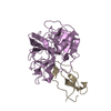
| ||||||||
|---|---|---|---|---|---|---|---|---|---|
| 1 |
| ||||||||
| Unit cell |
|
-Components
| #1: Protein | Mass: 25928.031 Da / Num. of mol.: 1 / Source method: isolated from a natural source / Source: (natural) |
|---|---|
| #2: Protein | Mass: 6014.225 Da / Num. of mol.: 1 Source method: isolated from a genetically manipulated source Source: (gene. exp.) Homo sapiens (human) / Organ: SKIN / References: UniProt: P19957 |
| #3: Water | ChemComp-HOH /  Mass: 18.015 Da / Num. of mol.: 157 / Source method: isolated from a natural source / Formula: H2O Mass: 18.015 Da / Num. of mol.: 157 / Source method: isolated from a natural source / Formula: H2O |
| Has protein modification | Y |
-Experimental details
-Experiment
| Experiment | Method: X-RAY DIFFRACTION / Number of used crystals: 1 |
|---|
- Sample preparation
| Crystal | Density Matthews: 2.05 Å3/Da / Density % sol: 40 % | |||||||||||||||
|---|---|---|---|---|---|---|---|---|---|---|---|---|---|---|---|---|
| Crystal grow | pH: 5.9 / Details: pH 5.9 | |||||||||||||||
| Crystal | *PLUS | |||||||||||||||
| Crystal grow | *PLUS Method: macroseeding | |||||||||||||||
| Components of the solutions | *PLUS
|
-Data collection
| Diffraction | Mean temperature: 293 K |
|---|---|
| Diffraction source | Wavelength: 1.5418 |
| Detector | Type: RIGAKU RAXIS IIC / Detector: IMAGE PLATE / Date: Dec 1, 1992 |
| Radiation | Monochromatic (M) / Laue (L): M / Scattering type: x-ray |
| Radiation wavelength | Wavelength: 1.5418 Å / Relative weight: 1 |
| Reflection | Num. obs: 15809 / % possible obs: 76.8 % / Observed criterion σ(I): 1 / Redundancy: 1.7 % / Rmerge(I) obs: 0.058 |
| Reflection | *PLUS Highest resolution: 1.9 Å / Num. measured all: 20575 |
- Processing
| Software |
| ||||||||||||||||||||||||||||||||||||||||||||||||||||||||||||||||||||||||||||||||||||
|---|---|---|---|---|---|---|---|---|---|---|---|---|---|---|---|---|---|---|---|---|---|---|---|---|---|---|---|---|---|---|---|---|---|---|---|---|---|---|---|---|---|---|---|---|---|---|---|---|---|---|---|---|---|---|---|---|---|---|---|---|---|---|---|---|---|---|---|---|---|---|---|---|---|---|---|---|---|---|---|---|---|---|---|---|---|
| Refinement | Resolution: 1.9→10 Å / σ(F): 1
| ||||||||||||||||||||||||||||||||||||||||||||||||||||||||||||||||||||||||||||||||||||
| Displacement parameters | Biso mean: 24.81 Å2 | ||||||||||||||||||||||||||||||||||||||||||||||||||||||||||||||||||||||||||||||||||||
| Refine analyze | Luzzati coordinate error obs: 0.21 Å / Luzzati d res low obs: 10 Å | ||||||||||||||||||||||||||||||||||||||||||||||||||||||||||||||||||||||||||||||||||||
| Refinement step | Cycle: LAST / Resolution: 1.9→10 Å
| ||||||||||||||||||||||||||||||||||||||||||||||||||||||||||||||||||||||||||||||||||||
| Refine LS restraints |
| ||||||||||||||||||||||||||||||||||||||||||||||||||||||||||||||||||||||||||||||||||||
| Software | *PLUS Name: PROFFT / Classification: refinement | ||||||||||||||||||||||||||||||||||||||||||||||||||||||||||||||||||||||||||||||||||||
| Refinement | *PLUS Num. reflection obs: 15659 / Rfactor obs: 0.197 | ||||||||||||||||||||||||||||||||||||||||||||||||||||||||||||||||||||||||||||||||||||
| Solvent computation | *PLUS | ||||||||||||||||||||||||||||||||||||||||||||||||||||||||||||||||||||||||||||||||||||
| Displacement parameters | *PLUS |
