2RMZ
 
 | |
5IOH
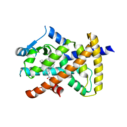
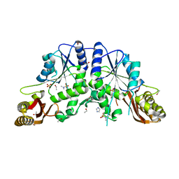
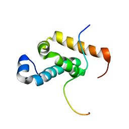
 | | RepoMan-PP1a (protein phosphatase 1, alpha isoform) holoenzyme complex | | 分子名称: | Cell division cycle-associated protein 2, Serine/threonine-protein phosphatase PP1-alpha catalytic subunit | | 著者 | Kumar, G.S, Peti, W, Page, R. | | 登録日 | 2016-03-08 | | 公開日 | 2016-10-05 | | 最終更新日 | 2023-09-27 | | 実験手法 | X-RAY DIFFRACTION (2.566 Å) | | 主引用文献 | The Ki-67 and RepoMan mitotic phosphatases assemble via an identical, yet novel mechanism.
Elife, 5, 2016
|
|
7ABT
 | |
2RB6
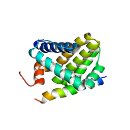
 | | X-Ray structure of the protein Q8EI81. Northeast Structural Genomics Consortium target SoR78A | | 分子名称: | Uncharacterized protein | | 著者 | Kuzin, A.P, Su, M, Seetharaman, J, Vorobiev, S.M, Wang, H, Mao, L, Cunningham, K, Xiao, R, Liu, J, Baran, M.C, Acton, T.B, Rost, B, Montelione, G.T, Hunt, J.F, Tong, L, Northeast Structural Genomics Consortium (NESG) | | 登録日 | 2007-09-18 | | 公開日 | 2007-10-23 | | 最終更新日 | 2017-10-25 | | 実験手法 | X-RAY DIFFRACTION (2.5 Å) | | 主引用文献 | X-Ray structure of the protein Q8EI81.
To be Published
|
|
6LVU
 | |
6LXL
 | |
5IK1
 | |
5IEB
 | |
2RMM
 | |
2RNE
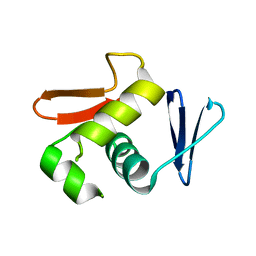
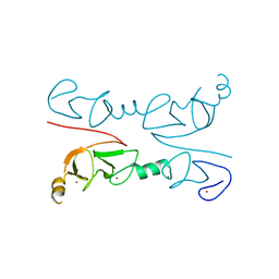
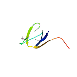
 | | Solution structure of the second RNA recognition motif (RRM) of TIA-1 | | 分子名称: | Tia1 protein | | 著者 | Takahashi, M, Kuwasako, K, Abe, C, Tsuda, K, Inoue, M, Terada, T, Shirouzu, M, Kobayashi, N, Kigawa, T, Taguchi, S, Guntert, P, Hayashizaki, Y, Tanaka, A, Muto, Y, Yokoyama, S. | | 登録日 | 2007-12-19 | | 公開日 | 2008-11-04 | | 最終更新日 | 2024-05-29 | | 実験手法 | SOLUTION NMR | | 主引用文献 | Solution structure of the second RNA recognition motif (RRM) domain of murine T cell intracellular antigen-1 (TIA-1) and its RNA recognition mode
Biochemistry, 47, 2008
|
|
6MD2
 | |
2ROC
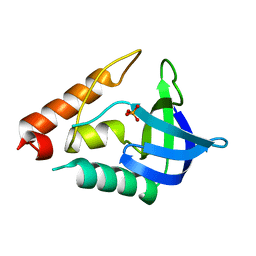
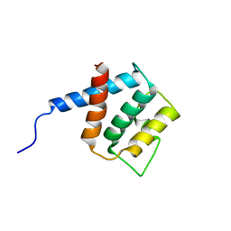
 | | Solution structure of Mcl-1 Complexed with Puma | | 分子名称: | Bcl-2-binding component 3, Induced myeloid leukemia cell differentiation protein Mcl-1 homolog | | 著者 | Day, C.L, Smits, C, Fan, F.C, Lee, E.F, Fairlie, W.D, Hinds, M.G. | | 登録日 | 2008-03-17 | | 公開日 | 2008-07-08 | | 最終更新日 | 2024-05-29 | | 実験手法 | SOLUTION NMR | | 主引用文献 | Structure of the BH3 Domains from the p53-Inducible BH3-Only Proteins Noxa and Puma in Complex with Mcl-1
J.Mol.Biol., 380, 2008
|
|
2RGT
 | | Crystal Structure of Lhx3 LIM domains 1 and 2 with the binding domain of Isl1 | | 分子名称: | Fusion of LIM/homeobox protein Lhx3, linker, Insulin gene enhancer protein ISL-1, ... | | 著者 | Bhati, M, Lee, M, Guss, J.M, Matthews, J.M. | | 登録日 | 2007-10-05 | | 公開日 | 2008-08-12 | | 最終更新日 | 2024-03-13 | | 実験手法 | X-RAY DIFFRACTION (2.05 Å) | | 主引用文献 | Implementing the LIM code: the structural basis for cell type-specific assembly of LIM-homeodomain complexes.
Embo J., 27, 2008
|
|
2RDF
 | |
4EFK
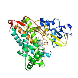
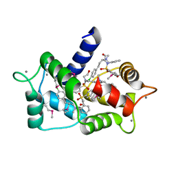
 | | Pantothenate synthetase in complex with N,N-DIMETHYLTHIOPHENE-3-SULFONAMIDE | | 分子名称: | ETHANOL, GLYCEROL, IMIDAZOLE, ... | | 著者 | Ciulli, A, Silvestre, H.L, Blundell, T.L, Abell, C. | | 登録日 | 2012-03-29 | | 公開日 | 2013-03-13 | | 最終更新日 | 2024-02-28 | | 実験手法 | X-RAY DIFFRACTION (1.7 Å) | | 主引用文献 | Integrated biophysical approach to fragment screening and validation for fragment-based lead discovery.
Proc.Natl.Acad.Sci.USA, 110, 2013
|
|
2RQ1
 | |
6M7H
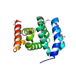
 | | Structure of calmodulin with KN93 | | 分子名称: | CALCIUM ION, Calmodulin-1, N-[2-[[[3-(4'-Chlorophenyl)-2-propenyl]methylamino]methyl]phenyl]-N-(2-hydroxyethyl)-4'-methoxybenzenesulfonamide | | 著者 | Damo, S.M, Pattanayek, R, Johnson, C.N. | | 登録日 | 2018-08-20 | | 公開日 | 2019-08-28 | | 最終更新日 | 2019-11-27 | | 実験手法 | X-RAY DIFFRACTION (1.6 Å) | | 主引用文献 | The CaMKII inhibitor KN93-calmodulin interaction and implications for calmodulin tuning of NaV1.5 and RyR2 function.
Cell Calcium, 82, 2019
|
|
5IOI
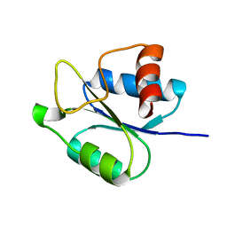
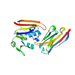
 | | X-RAY STRUCTURE OF THE N-TERMINAL DOMAIN OF HUMAN DOUBLECORTIN | | 分子名称: | Neuronal migration protein doublecortin | | 著者 | Ruf, A, Benz, J, Burger, D, D'Arcy, B, Debulpaep, M, Di Lello, P, Fry, D, Huber, W, Kremer, T, Laeremans, T, Matile, H, Ross, A, Rudolph, M.G, Rufer, A.C, Sharma, A, Steinmetz, M.O, Steyaert, J, Schoch, G, Stihle, M, Thoma, R. | | 登録日 | 2016-03-08 | | 公開日 | 2016-03-23 | | 最終更新日 | 2024-01-10 | | 実験手法 | X-RAY DIFFRACTION (2.4 Å) | | 主引用文献 | Crystal Structures of the Human Doublecortin C- and N-terminal Domains in Complex with Specific Antibodies.
J.Biol.Chem., 291, 2016
|
|
2RD1
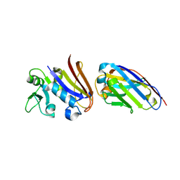
 | | X-Ray structure of the protein Q7CQI7. Northeast Structural Genomics Consortium target StR87A | | 分子名称: | Putative outer membrane lipoprotein | | 著者 | Kuzin, A.P, Su, M, Seetharaman, J, Vorobiev, S.M, Wang, D, Fang, Y, Owens, L, Mao, L.-C, Xiao, R, Liu, J, Baran, M.C, Acton, T.B, Rost, B, Montelione, G.T, Hunt, J.F, Tong, L, Northeast Structural Genomics Consortium (NESG) | | 登録日 | 2007-09-20 | | 公開日 | 2007-10-09 | | 最終更新日 | 2018-01-24 | | 実験手法 | X-RAY DIFFRACTION (2.3 Å) | | 主引用文献 | X-Ray structure of the protein Q7CQI7.
To be Published
|
|
2RQH
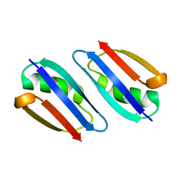
 | | Structure of GSPT1/ERF3A-PABC | | 分子名称: | G1 to S phase transition 1, Polyadenylate-binding protein 1 | | 著者 | Osawa, M, Nakanishi, T, Hosoda, N, Uchida, S, Hoshino, T, Katada, I, Shimada, I. | | 登録日 | 2009-05-08 | | 公開日 | 2010-05-26 | | 最終更新日 | 2024-05-01 | | 実験手法 | SOLUTION NMR | | 主引用文献 | Eukaryotic Translation Termination Factor Gspt/Erf3 Recognizes Pabp with Chemical Exchange Using Two Overlapping Motifs
To be Published
|
|
2RG8
 | |
5IM8
 | |
4EIG
 | | CA1698 camel antibody fragment in complex with DHFR | | 分子名称: | CA1698 camel antibody fragment, Dihydrofolate reductase | | 著者 | Oyen, D, Srinivasan, V. | | 登録日 | 2012-04-05 | | 公開日 | 2013-04-24 | | 最終更新日 | 2017-11-15 | | 実験手法 | X-RAY DIFFRACTION (2.5 Å) | | 主引用文献 | Mechanistic analysis of allosteric and non-allosteric effects arising from nanobody binding to two epitopes of the dihyrofolate reductase of Escherichia coli.
Biochim.Biophys.Acta, 1834, 2013
|
|
4EIZ
 | | Structure of Nb113 bound to apoDHFR | | 分子名称: | Dihydrofolate reductase, Nb113 Camel antibody fragment | | 著者 | Oyen, D, Srinivasan, V. | | 登録日 | 2012-04-06 | | 公開日 | 2013-04-24 | | 最終更新日 | 2013-10-30 | | 実験手法 | X-RAY DIFFRACTION (2.2 Å) | | 主引用文献 | Mechanistic analysis of allosteric and non-allosteric effects arising from nanobody binding to two epitopes of the dihyrofolate reductase of Escherichia coli.
Biochim.Biophys.Acta, 1834, 2013
|
|
6MG5
 | | Structure of full-length human lambda-6A light chain JTO in complex with coumarin 1 | | 分子名称: | 7-(diethylamino)-4-methyl-2H-1-benzopyran-2-one, Light chain JTO, PHOSPHATE ION | | 著者 | Morgan, G.J, Yan, N.L, Mortenson, D.E, Stanfield, R.L, Wilson, I.A, Kelly, J.W. | | 登録日 | 2018-09-12 | | 公開日 | 2019-04-10 | | 最終更新日 | 2019-12-25 | | 実験手法 | X-RAY DIFFRACTION (1.8 Å) | | 主引用文献 | Stabilization of amyloidogenic immunoglobulin light chains by small molecules.
Proc.Natl.Acad.Sci.USA, 116, 2019
|
|


