7C1D
 
 | |
7E0F
 | |
7QEU
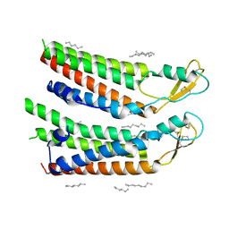
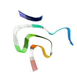
 | | human Connexin 26 at 55mmHg PCO2, pH7.4: two masked subunits, class B | | 分子名称: | DODECYL-BETA-D-MALTOSIDE, Gap junction beta-2 protein, PHOSPHATIDYLETHANOLAMINE | | 著者 | Brotherton, D.H, Cameron, A.D, Savva, C.G, Ragan, T.J. | | 登録日 | 2021-12-03 | | 公開日 | 2022-06-15 | | 実験手法 | ELECTRON MICROSCOPY (2.7 Å) | | 主引用文献 | Conformational changes and CO 2 -induced channel gating in connexin26.
Structure, 30, 2022
|
|
7QEV
 | | human Connexin 26 at 55mm Hg PCO2, pH7.4:two masked subunits, class D | | 分子名称: | DODECYL-BETA-D-MALTOSIDE, Gap junction beta-2 protein, PHOSPHATIDYLETHANOLAMINE | | 著者 | Brotherton, D.H, Cameron, A.D, Savva, C.G, Ragan, T.J. | | 登録日 | 2021-12-03 | | 公開日 | 2022-06-15 | | 実験手法 | ELECTRON MICROSCOPY (2.9 Å) | | 主引用文献 | Conformational changes and CO 2 -induced channel gating in connexin26.
Structure, 30, 2022
|
|
7QES
 | | human Connexin 26 at 55mm Hg PCO2, pH7.4: two masked subunits, class A | | 分子名称: | DODECYL-BETA-D-MALTOSIDE, Gap junction beta-2 protein, PHOSPHATIDYLETHANOLAMINE | | 著者 | Brotherton, D.H, Cameron, A.D, Savva, C.G, Ragan, T.J. | | 登録日 | 2021-12-03 | | 公開日 | 2022-06-15 | | 実験手法 | ELECTRON MICROSCOPY (2.6 Å) | | 主引用文献 | Conformational changes and CO 2 -induced channel gating in connexin26.
Structure, 30, 2022
|
|
2WGO
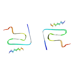
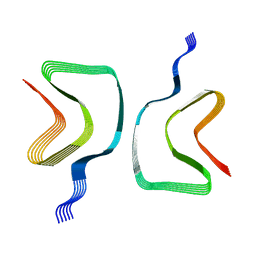
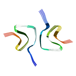
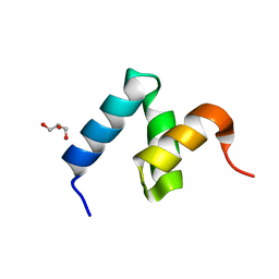
 | | Structure of ranaspumin-2, a surfactant protein from the foam nests of a tropical frog | | 分子名称: | RANASPUMIN-2 | | 著者 | Mackenzie, C.D, Smith, B.O, Kennedy, M.W, Cooper, A. | | 登録日 | 2009-04-21 | | 公開日 | 2009-06-23 | | 最終更新日 | 2011-07-13 | | 実験手法 | SOLUTION NMR | | 主引用文献 | Ranaspumin-2: Structure and Function of a Surfactant Protein from the Foam Nests of a Tropical Frog.
Biophys.J., 96, 2009
|
|
8JEX
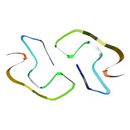
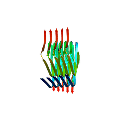
 | | Cryo-EM structure of alpha-synuclein gS87 fibril | | 分子名称: | 2-acetamido-2-deoxy-beta-D-glucopyranose, Alpha-synuclein, unverified amino acid chain | | 著者 | Xia, W.C, Sun, Y.P, Liu, C. | | 登録日 | 2023-05-16 | | 公開日 | 2024-04-17 | | 最終更新日 | 2024-05-08 | | 実験手法 | ELECTRON MICROSCOPY (3.1 Å) | | 主引用文献 | Phosphorylation and O-GlcNAcylation at the same alpha-synuclein site generate distinct fibril structures.
Nat Commun, 15, 2024
|
|
8JEY
 | |
8PK2
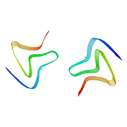
 | | Cryo EM structure of the type 1m polymorph of alpha-synuclein | | 分子名称: | Alpha-synuclein | | 著者 | Frey, L, Qureshi, B.M, Kwiatkowski, W, Rhyner, D, Greenwald, J, Riek, R. | | 登録日 | 2023-06-24 | | 公開日 | 2024-05-29 | | 実験手法 | ELECTRON MICROSCOPY (3.26 Å) | | 主引用文献 | On the pH-dependence of alpha-synuclein amyloid polymorphism and the role of secondary nucleation in seed-based amyloid propagation
Elife, 2023
|
|
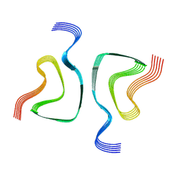
8PIX
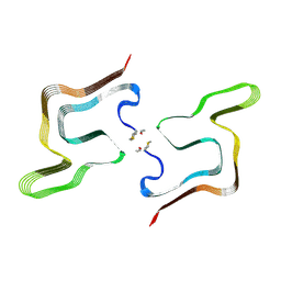
 | | Cryo EM structure of the type 3C polymorph of alpha-synuclein at low pH. | | 分子名称: | Alpha-synuclein | | 著者 | Frey, L, Qureshi, B.M, Kwiatkowski, W, Rhyner, D, Greenwald, J, Riek, R. | | 登録日 | 2023-06-22 | | 公開日 | 2024-05-29 | | 実験手法 | ELECTRON MICROSCOPY (3.41 Å) | | 主引用文献 | On the pH-dependence of alpha-synuclein amyloid polymorphism and the role of secondary nucleation in seed-based amyloid propagation
Elife, 2023
|
|
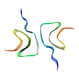
8PJO
 | | Cryo EM structure of the type 3D polymorph of alpha-synuclein E46K mutant at low pH. | | 分子名称: | Alpha-synuclein, CHLORIDE ION | | 著者 | Frey, L, Qureshi, B.M, Kwiatkowski, W, Rhyner, D, Greenwald, J, Riek, R. | | 登録日 | 2023-06-23 | | 公開日 | 2024-05-29 | | 実験手法 | ELECTRON MICROSCOPY (2.31 Å) | | 主引用文献 | On the pH-dependence of alpha-synuclein amyloid polymorphism and the role of secondary nucleation in seed-based amyloid propagation
Elife, 2023
|
|
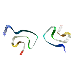
8PK4
 | | Cryo EM structure of the type 5A polymorph of alpha-synuclein. | | 分子名称: | Alpha-synuclein | | 著者 | Frey, L, Qureshi, B.M, Kwiatkowski, W, Rhyner, D, Greenwald, J, Riek, R. | | 登録日 | 2023-06-24 | | 公開日 | 2024-05-29 | | 実験手法 | ELECTRON MICROSCOPY (3.3 Å) | | 主引用文献 | On the pH-dependence of alpha-synuclein amyloid polymorphism and the role of secondary nucleation in seed-based amyloid propagation
Elife, 2023
|
|
8OQI
 | | Cryo-EM structure of the wild-type alpha-synuclein fibril. | | 分子名称: | Alpha-synuclein | | 著者 | Pesch, V, Reithofer, S, Ma, L, Flores-Fernandez, J.M, Oezduezenciler, P, Busch, Y, Lien, Y, Rudtke, O, Frieg, B, Schroeder, G.F, Wille, H, Tamgueney, G. | | 登録日 | 2023-04-12 | | 公開日 | 2024-03-13 | | 最終更新日 | 2024-05-15 | | 実験手法 | ELECTRON MICROSCOPY (3.1 Å) | | 主引用文献 | Vaccination with structurally adapted fungal protein fibrils induces immunity to Parkinson's disease.
Brain, 147, 2024
|
|
5YHJ
 | | Cytochrome P450EX alpha (CYP152N1) wild-type with myristic acid | | 分子名称: | Cytochrome P450, MYRISTIC ACID, PROTOPORPHYRIN IX CONTAINING FE | | 著者 | Onoda, H, Shoji, O, Suzuki, K, Sugimoto, H, Shiro, Y, Watanabe, Y. | | 登録日 | 2017-09-28 | | 公開日 | 2017-12-06 | | 最終更新日 | 2024-03-27 | | 実験手法 | X-RAY DIFFRACTION (2.3 Å) | | 主引用文献 | Alpha-Oxidative Decarboxylation of Fatty Acids Catalysed by Cytochrome P450 Peroxygenases Yielding Shorter-Alkyl-Chain Fatty Acids
Catalysis Science And Technology, 2017
|
|
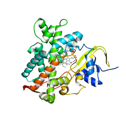
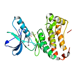
4P5Q
 | | Human EphA3 Kinase domain in complex with quinoxaline derivatives | | 分子名称: | 2-amino-1-(2-chlorophenyl)-N-(3-ethoxypropyl)-1H-pyrrolo[2,3-b]quinoxaline-3-carboxamide, Ephrin type-A receptor 3 | | 著者 | Dong, J, Caflisch, A. | | 登録日 | 2014-03-19 | | 公開日 | 2014-08-13 | | 最終更新日 | 2023-12-20 | | 実験手法 | X-RAY DIFFRACTION (1.35 Å) | | 主引用文献 | Pyrrolo[3,2-b]quinoxaline Derivatives as Types I1/2 and II Eph Tyrosine Kinase Inhibitors: Structure-Based Design, Synthesis, and in Vivo Validation.
J.Med.Chem., 57, 2014
|
|
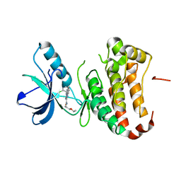
4P4C
 | | Human EphA3 Kinase domain in complex with quinoxaline derivatives | | 分子名称: | 2-amino-1-(3-methoxyphenyl)-1H-pyrrolo[2,3-b]quinoxaline-3-carboxamide, EPH receptor A3 | | 著者 | Dong, J, Caflisch, A. | | 登録日 | 2014-03-12 | | 公開日 | 2014-08-13 | | 最終更新日 | 2023-09-27 | | 実験手法 | X-RAY DIFFRACTION (1.599 Å) | | 主引用文献 | Pyrrolo[3,2-b]quinoxaline Derivatives as Types I1/2 and II Eph Tyrosine Kinase Inhibitors: Structure-Based Design, Synthesis, and in Vivo Validation.
J.Med.Chem., 57, 2014
|
|
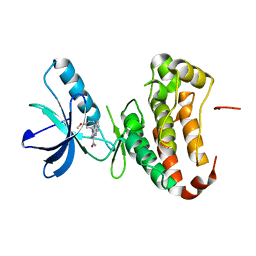
4P5Z
 | | Human EphA3 Kinase domain in complex with quinoxaline derivatives | | 分子名称: | 2-amino-1-[4-({[3-(trifluoromethyl)phenyl]carbamoyl}amino)phenyl]-1H-pyrrolo[2,3-b]quinoxaline-3-carboxamide, Ephrin type-A receptor 3 | | 著者 | Dong, J, Caflisch, A. | | 登録日 | 2014-03-20 | | 公開日 | 2014-08-13 | | 最終更新日 | 2023-12-20 | | 実験手法 | X-RAY DIFFRACTION (2.002 Å) | | 主引用文献 | Pyrrolo[3,2-b]quinoxaline Derivatives as Types I1/2 and II Eph Tyrosine Kinase Inhibitors: Structure-Based Design, Synthesis, and in Vivo Validation.
J.Med.Chem., 57, 2014
|
|
6OSM
 | |
6OSJ
 | |
6PES
 | | Cryo-EM structure of alpha-synuclein H50Q Wide Fibril | | 分子名称: | Alpha-synuclein | | 著者 | Boyer, D.R, Li, B, Sawaya, M.R, Jiang, L, Eisenberg, D.S. | | 登録日 | 2019-06-20 | | 公開日 | 2019-11-27 | | 最終更新日 | 2024-03-20 | | 実験手法 | ELECTRON MICROSCOPY (3.6 Å) | | 主引用文献 | Structures of fibrils formed by alpha-synuclein hereditary disease mutant H50Q reveal new polymorphs.
Nat.Struct.Mol.Biol., 26, 2019
|
|
6OSL
 | |
7KWO
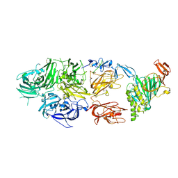 | | rFVIIIFc-VWF-XTEN (BIVV001) | | 分子名称: | 2-acetamido-2-deoxy-beta-D-glucopyranose, CALCIUM ION, COPPER (II) ION, ... | | 著者 | Fuller, J.R, Batchelor, J.D. | | 登録日 | 2020-12-01 | | 公開日 | 2021-03-03 | | 最終更新日 | 2021-06-09 | | 実験手法 | ELECTRON MICROSCOPY (2.9 Å) | | 主引用文献 | Molecular determinants of the factor VIII/von Willebrand factor complex revealed by BIVV001 cryo-electron microscopy.
Blood, 137, 2021
|
|
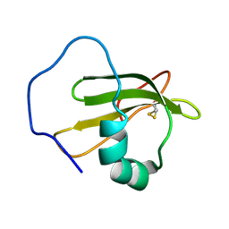
6W2G
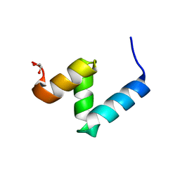 | | Crystal Structure of Y188G Variant of the Internal UBA Domain of HHR23A in Monoclinic Unit Cell | | 分子名称: | 1,2-ETHANEDIOL, UV excision repair protein RAD23 homolog A | | 著者 | Bowler, B.E, Zeng, B, Becht, D.C, Rothfuss, M, Sprang, S.R, Mou, T.-C. | | 登録日 | 2020-03-05 | | 公開日 | 2021-03-10 | | 最終更新日 | 2024-04-03 | | 実験手法 | X-RAY DIFFRACTION (1.1 Å) | | 主引用文献 | High-Accuracy Prediction of Stabilizing Surface Mutations to the Three-Helix Bundle, UBA(1), with EmCAST.
J.Am.Chem.Soc., 145, 2023
|
|
6W2H
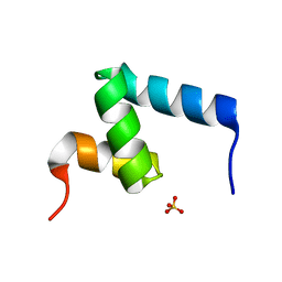 | | Crystal Structure of the Internal UBA Domain of HHR23A | | 分子名称: | SULFATE ION, UV excision repair protein RAD23 homolog A | | 著者 | Bowler, B.E, Zeng, B, Becht, D.C, Rothfuss, M, Sprang, S.R, Mou, T.-C. | | 登録日 | 2020-03-05 | | 公開日 | 2021-03-10 | | 最終更新日 | 2024-04-03 | | 実験手法 | X-RAY DIFFRACTION (1.6 Å) | | 主引用文献 | Residual Structure in the Denatured State of the Fast-Folding UBA(1) Domain from the Human DNA Excision Repair Protein HHR23A.
Biochemistry, 61, 2022
|
|
6W2I
 | | Crystal Structure of Y188G Variant of the Internal UBA Domain of HHR23A | | 分子名称: | GLYCEROL, UV excision repair protein RAD23 homolog A | | 著者 | Bowler, B.E, Zeng, B, Becht, D.C, Rothfuss, M, Sprang, S.R, Mou, T.-C. | | 登録日 | 2020-03-05 | | 公開日 | 2021-03-10 | | 最終更新日 | 2023-11-01 | | 実験手法 | X-RAY DIFFRACTION (1.45 Å) | | 主引用文献 | High-Accuracy Prediction of Stabilizing Surface Mutations to the Three-Helix Bundle, UBA(1), with EmCAST.
J.Am.Chem.Soc., 145, 2023
|
|


