6D8X
 
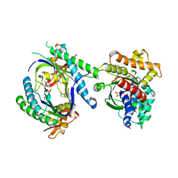
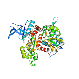
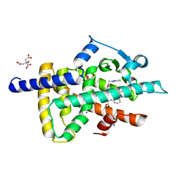 | | PPAR gamma LBD complexed with the agonist GW1929 | | 分子名称: | (2~{S})-3-[4-[2-[methyl(pyridin-2-yl)amino]ethoxy]phenyl]-2-[[2-(phenylcarbonyl)phenyl]amino]propanoic acid, CITRATE ANION, GLYCEROL, ... | | 著者 | Mou, T.C, Chrisman, I.M, Hughes, T.S, Sprang, S.R. | | 登録日 | 2018-04-27 | | 公開日 | 2019-05-01 | | 最終更新日 | 2023-10-04 | | 実験手法 | X-RAY DIFFRACTION (1.9 Å) | | 主引用文献 | PPAR gamma LBD complexed with the agonist GW1929
To Be Published
|
|
6DBH
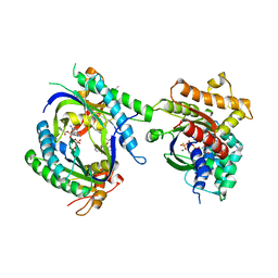
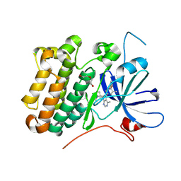
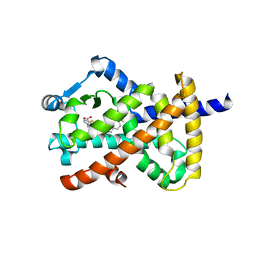 | | Crystal structure of PPAR gamma in complex with NMP422 | | 分子名称: | 4-{2-[(2,3-dioxo-1-pentyl-2,3-dihydro-1H-indol-5-yl)sulfanyl]ethyl}benzoic acid, CHLORIDE ION, Peroxisome proliferator-activated receptor gamma | | 著者 | Mou, T.C, Chrisman, I.M, Hughes, T.S, Sprang, S.R. | | 登録日 | 2018-05-03 | | 公開日 | 2019-05-08 | | 最終更新日 | 2023-10-04 | | 実験手法 | X-RAY DIFFRACTION (2.597 Å) | | 主引用文献 | Crystal structure of PPAR gamma in complex with NMP422
To Be Published
|
|
6DCU
 | |
6D94
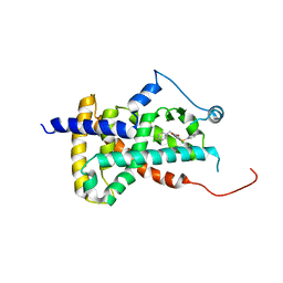
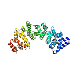
 | | Crystal structure of PPAR gamma in complex with Mediator of RNA polymerase II transcription subunit 1 | | 分子名称: | (2~{S})-3-[4-[2-[methyl(pyridin-2-yl)amino]ethoxy]phenyl]-2-[[2-(phenylcarbonyl)phenyl]amino]propanoic acid, Mediator of RNA polymerase II transcription subunit 1, Peroxisome proliferator-activated receptor gamma | | 著者 | Mou, T.C, Chrisman, I.M, Hughes, T.S, Sprang, S.R. | | 登録日 | 2018-04-27 | | 公開日 | 2019-05-01 | | 最終更新日 | 2023-10-25 | | 実験手法 | X-RAY DIFFRACTION (1.9 Å) | | 主引用文献 | A structural mechanism of nuclear receptor biased agonism.
Proc.Natl.Acad.Sci.USA, 119, 2022
|
|
1U0H
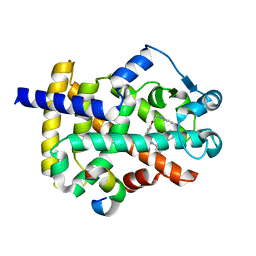
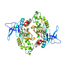
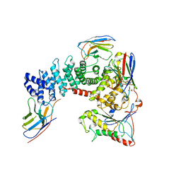
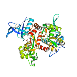
 | | STRUCTURAL BASIS FOR THE INHIBITION OF MAMMALIAN ADENYLYL CYCLASE BY MANT-GTP | | 分子名称: | 3'-O-(N-METHYLANTHRANILOYL)-GUANOSINE-5'-TRIPHOSPHATE, 5'-GUANOSINE-DIPHOSPHATE-MONOTHIOPHOSPHATE, Adenylate cyclase, ... | | 著者 | Mou, T.C, Gille, A, Seifert, R.J, Sprang, S.R. | | 登録日 | 2004-07-13 | | 公開日 | 2004-12-14 | | 最終更新日 | 2023-08-23 | | 実験手法 | X-RAY DIFFRACTION (2.9 Å) | | 主引用文献 | Structural basis for the inhibition of mammalian membrane adenylyl cyclase by 2 '(3')-O-(N-Methylanthraniloyl)-guanosine 5 '-triphosphate.
J.Biol.Chem., 280, 2005
|
|
1TL7
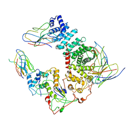
 | | Complex Of Gs- With The Catalytic Domains Of Mammalian Adenylyl Cyclase: Complex With 2'(3')-O-(N-methylanthraniloyl)-guanosine 5'-triphosphate and Mn | | 分子名称: | 3'-O-(N-METHYLANTHRANILOYL)-GUANOSINE-5'-TRIPHOSPHATE, 5'-GUANOSINE-DIPHOSPHATE-MONOTHIOPHOSPHATE, Adenylate cyclase, ... | | 著者 | Mou, T.C, Gille, A, Seifert, R.J, Sprang, S.R. | | 登録日 | 2004-06-09 | | 公開日 | 2004-12-14 | | 最終更新日 | 2023-08-23 | | 実験手法 | X-RAY DIFFRACTION (2.8 Å) | | 主引用文献 | Structural basis for the inhibition of mammalian membrane adenylyl cyclase by 2 '(3')-O-(N-Methylanthraniloyl)-guanosine 5 '-triphosphate.
J.Biol.Chem., 280, 2005
|
|
6XAZ
 | |
6XAX
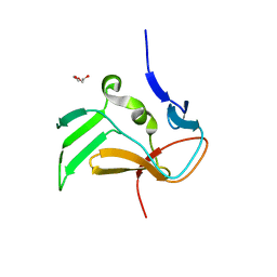
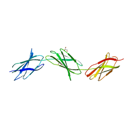
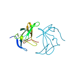
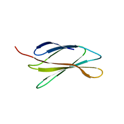
 | | Structure of a fragment of human fibronectin containing the 11th type III domain, extra domain A, and the 12th type III domain | | 分子名称: | Fibronectin, GLYCEROL | | 著者 | Mou, T.C, Nepomuceno, P.A, Sprang, S.R, Briknarova, K. | | 登録日 | 2020-06-04 | | 公開日 | 2021-06-09 | | 最終更新日 | 2023-10-18 | | 実験手法 | X-RAY DIFFRACTION (2.4 Å) | | 主引用文献 | Fragment of human fibronectin containing the 11th type III domain, extra domain A, and 12th type III domain
To Be Published
|
|
6XAY
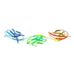
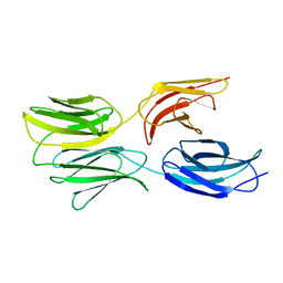
 | | Structure of a fragment of human fibronectin containing the 10th, 11th and 12th type III domains | | 分子名称: | Fibronectin, GLYCEROL | | 著者 | Mou, T.C, Nepomuceno, P.A, Sprang, S.R, Briknarova, K. | | 登録日 | 2020-06-05 | | 公開日 | 2021-06-09 | | 最終更新日 | 2023-10-18 | | 実験手法 | X-RAY DIFFRACTION (2.48 Å) | | 主引用文献 | Fragment of human fibronectin containing the 10th, 11th and 12th type III domains
To Be Published
|
|
6ODL
 | | Crystal structure of GluN2A agonist binding domain with 4-butyl-(S)-CCG-IV | | 分子名称: | (1S,2R)-2-[(S)-amino(carboxy)methyl]-1-butylcyclopropane-1-carboxylic acid, Glutamate receptor ionotropic, NMDA 2A,Glutamate receptor ionotropic, ... | | 著者 | Mou, T.C, Clausen, R.P, Sprang, S.R, Hansen, K.B. | | 登録日 | 2019-03-26 | | 公開日 | 2020-04-01 | | 最終更新日 | 2023-10-11 | | 実験手法 | X-RAY DIFFRACTION (2.3 Å) | | 主引用文献 | Stereoselective synthesis of novel 2'-(S)-CCG-IV analogues as potent NMDA receptor agonists.
Eur.J.Med.Chem., 212, 2021
|
|
5DFS
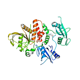
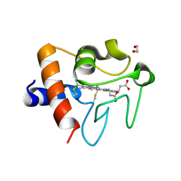 | | Crystal structure of Spider Monkey Cytochrome C at 1.15 Angstrom | | 分子名称: | 1,2-ETHANEDIOL, CHLORIDE ION, Cytochrome c, ... | | 著者 | MOU, T.C, McClelland, L.J, Jeakins-Cooley, M.E, Goldes, M.E, SPRANG, S.R, BOWLER, B.E. | | 登録日 | 2015-08-27 | | 公開日 | 2016-03-02 | | 最終更新日 | 2023-09-27 | | 実験手法 | X-RAY DIFFRACTION (1.15 Å) | | 主引用文献 | Disruption of a hydrogen bond network in human versus spider monkey cytochrome c affects heme crevice stability.
J.Inorg.Biochem., 158, 2016
|
|
5TY3
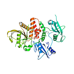
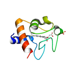 | | Crystal structure of K72A variant of Human Cytochrome c | | 分子名称: | Cytochrome c, HEME C, SULFATE ION | | 著者 | Mou, T.C, Nold, S.M, Lei, H, Sprang, S.R, Bowler, B.E. | | 登録日 | 2016-11-18 | | 公開日 | 2017-09-06 | | 最終更新日 | 2023-10-04 | | 実験手法 | X-RAY DIFFRACTION (1.25 Å) | | 主引用文献 | Effect of a K72A Mutation on the Structure, Stability, Dynamics, and Peroxidase Activity of Human Cytochrome c.
Biochemistry, 56, 2017
|
|
6TYL
 | | Crystal structure of mammalian Ric-8A:Galpha(i):nanobody complex | | 分子名称: | Guanine nucleotide-binding protein G(i) subunit alpha-1, Nanobody A, Nanobody B, ... | | 著者 | Mou, T.C, McClelland, L, Yates-Hansen, C, Sprang, S.R. | | 登録日 | 2019-08-09 | | 公開日 | 2020-03-11 | | 最終更新日 | 2023-10-11 | | 実験手法 | X-RAY DIFFRACTION (3.3 Å) | | 主引用文献 | Structure of the G protein chaperone and guanine nucleotide exchange factor Ric-8A bound to G alpha i1.
Nat Commun, 11, 2020
|
|
6UKT
 | | Cryo-EM structure of mammalian Ric-8A:Galpha(i):nanobody complex | | 分子名称: | Guanine nucleotide-binding protein G(i) subunit alpha-1, NB8109, NB8117, ... | | 著者 | Mou, T.C, Zhang, K, Johnston, J.D, Chiu, W, Sprang, S.R. | | 登録日 | 2019-10-05 | | 公開日 | 2020-03-11 | | 実験手法 | ELECTRON MICROSCOPY (3.87 Å) | | 主引用文献 | Structure of the G protein chaperone and guanine nucleotide exchange factor Ric-8A bound to G alpha i1.
Nat Commun, 11, 2020
|
|
8U7X
 | |
8U7W
 | |
6NMJ
 | | Crystal Structure of Rat Ric-8A G alpha binding domain, "Paratone-N Immersed" | | 分子名称: | Resistance to inhibitors of cholinesterase 8 homolog A (C. elegans) | | 著者 | Zeng, B, Mou, T.C, Sprang, S.R. | | 登録日 | 2019-01-11 | | 公開日 | 2019-06-26 | | 最終更新日 | 2023-10-11 | | 実験手法 | X-RAY DIFFRACTION (2.3 Å) | | 主引用文献 | Structure, Function, and Dynamics of the G alpha Binding Domain of Ric-8A.
Structure, 27, 2019
|
|
6AU4
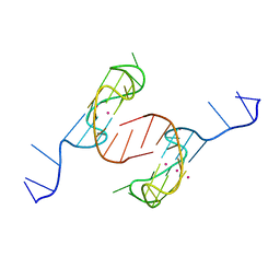 | | Crystal structure of the major quadruplex formed in the human c-MYC promoter | | 分子名称: | DNA (5'-D(*TP*GP*AP*GP*GP*GP*TP*GP*GP*GP*TP*AP*GP*GP*GP*TP*GP*GP*GP*TP*AP*A)-3'), POTASSIUM ION | | 著者 | Stump, S, Mou, T.C, Sprang, S.R, Natale, N.R, Beall, H.D. | | 登録日 | 2017-08-30 | | 公開日 | 2018-09-12 | | 最終更新日 | 2023-10-04 | | 実験手法 | X-RAY DIFFRACTION (2.35 Å) | | 主引用文献 | Crystal structure of the major quadruplex formed in the promoter region of the human c-MYC oncogene.
PLoS ONE, 13, 2018
|
|
6XAU
 | |
6OVD
 | | Crystal structure of GluN1/GluN2A NMDA receptor agonist binding domains with glycine and antagonist, 3-ethylphenyl-ACEPC | | 分子名称: | (3S,5S)-5-[(2R)-2-amino-2-carboxyethyl]-1-(3-ethylphenyl)pyrazolidine-3-carboxylic acid, GLYCINE, Glutamate receptor ionotropic, ... | | 著者 | Syrenne, J.T, Mou, T.C, Tamborini, L, Pinto, A, Sprang, S.R, Hansen, K.B. | | 登録日 | 2019-05-07 | | 公開日 | 2020-05-13 | | 最終更新日 | 2023-10-11 | | 実験手法 | X-RAY DIFFRACTION (2.102 Å) | | 主引用文献 | Crystal structure of GluN1/GluN2A NMDA receptor agonist binding domains with glycine and antagonist, 3-ethylphenyl-ACEPC
To Be Published
|
|
6OVE
 | | Crystal structure of GluN1/GluN2A NMDA receptor agonist binding domains with glycine and antagonist, 4-propylphenyl-ACEPC | | 分子名称: | (3R,5S)-5-[(2R)-2-amino-2-carboxyethyl]-1-(4-propylphenyl)pyrazolidine-3-carboxylic acid, GLYCINE, Glutamate receptor ionotropic, ... | | 著者 | Syrenne, J.T, Mou, T.C, Tamborini, L, Pinto, A, Sprang, S.R, Hansen, K.B. | | 登録日 | 2019-05-07 | | 公開日 | 2020-05-13 | | 最終更新日 | 2023-10-11 | | 実験手法 | X-RAY DIFFRACTION (2 Å) | | 主引用文献 | Crystal structure of GluN1/GluN2A NMDA receptor agonist binding domains with glycine and antagonist, 3-ethylphenyl-ACEPC
To Be Published
|
|
8U8X
 | | crystal structure of the receptor tyrosine kinase Human HER2 (ERBB2) YVMA mutant kinase domain in complex with inhibitor compound 27 | | 分子名称: | 1-{(1R,3r,5S)-3-[(3M)-4-methyl-3-{3-methyl-4-[(1-methyl-1H-benzimidazol-5-yl)oxy]phenyl}-1H-pyrazolo[3,4-d]pyrimidin-1-yl]-8-azabicyclo[3.2.1]octan-8-yl}propan-1-one, CHLORIDE ION, DI(HYDROXYETHYL)ETHER, ... | | 著者 | Wang, J, Mou, T.C. | | 登録日 | 2023-09-18 | | 公開日 | 2024-06-12 | | 最終更新日 | 2024-06-26 | | 実験手法 | X-RAY DIFFRACTION (1.69 Å) | | 主引用文献 | Discovery of Potent and Selective Covalent Inhibitors of HER2 WT and HER2 YVMA .
J.Med.Chem., 67, 2024
|
|
5DFT
 | |
6MFA
 | |
6MSV
 | |


