8BG9
 
 | | Murine amyloid-beta filaments with the Arctic mutation (E22G) from APP(NL-G-F) mouse brains | ABeta | | 分子名称: | Amyloid-beta protein 40 | | 著者 | Yang, Y, Zhang, W.J, Murzin, A.G, Schweighauser, M, Huang, M, Lovestam, S.K.A, Peak-Chew, S.Y, Macdonald, J, Lavenir, I, Ghetti, B, Graff, C, Kumar, A, Nordber, A, Goedert, M, Scheres, S.H.W. | | 登録日 | 2022-10-27 | | 公開日 | 2023-01-18 | | 最終更新日 | 2023-02-22 | | 実験手法 | ELECTRON MICROSCOPY (3.5 Å) | | 主引用文献 | Cryo-EM structures of amyloid-beta filaments with the Arctic mutation (E22G) from human and mouse brains.
Acta Neuropathol, 145, 2023
|
|
8BG0
 | | Amyloid-beta tetrameric filaments with the Arctic mutation (E22G) from Alzheimer's disease brains | ABeta40 | | 分子名称: | Amyloid-beta precursor protein | | 著者 | Yang, Y, Zhang, W.J, Murzin, A.G, Schweighauser, M, Huang, M, Lovestam, S.K.A, Peak-Chew, S.Y, Macdonald, J, Lavenir, I, Ghetti, B, Graff, C, Kumar, A, Nordber, A, Goedert, M, Scheres, S.H.W. | | 登録日 | 2022-10-27 | | 公開日 | 2023-01-18 | | 最終更新日 | 2023-02-22 | | 実験手法 | ELECTRON MICROSCOPY (1.9 Å) | | 主引用文献 | Cryo-EM structures of amyloid-beta filaments with the Arctic mutation (E22G) from human and mouse brains.
Acta Neuropathol, 145, 2023
|
|
8BFZ
 | | Amyloid-beta 42 filaments extracted from the human brain with Arctic mutation (E22G) of Alzheimer's disease | ABeta42 | | 分子名称: | Amyloid-beta precursor protein | | 著者 | Yang, Y, Zhang, W.J, Murzin, A.G, Schweighauser, M, Huang, M, Lovestam, S.K.A, Peak-Chew, S.Y, Macdonald, J, Lavenir, I, Ghetti, B, Graff, C, Kumar, A, Nordberg, A, Goedert, M, Scheres, S.H.W. | | 登録日 | 2022-10-27 | | 公開日 | 2023-01-18 | | 最終更新日 | 2024-01-31 | | 実験手法 | ELECTRON MICROSCOPY (2.8 Å) | | 主引用文献 | Cryo-EM structures of amyloid-beta filaments with the Arctic mutation (E22G) from human and mouse brains.
Acta Neuropathol, 145, 2023
|
|
4X1N
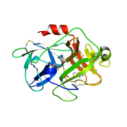
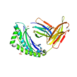
 | | The crystal structure of mupain-1-16 in complex with murinised human uPA at pH7.4 | | 分子名称: | Urokinase-type plasminogen activator, mupain-1-16, piperidine-1-carboximidamide | | 著者 | Jiang, L, Zhao, B, Xu, P, Andreasen, P, Huang, M. | | 登録日 | 2014-11-25 | | 公開日 | 2015-03-25 | | 最終更新日 | 2023-11-08 | | 実験手法 | X-RAY DIFFRACTION (1.8 Å) | | 主引用文献 | A cyclic peptidic serine protease inhibitor: increasing affinity by increasing peptide flexibility.
Plos One, 9, 2014
|
|
4X0W
 | | The crystal structure of mupain-1-17 in complex with murinised human uPA | | 分子名称: | SULFATE ION, Urokinase-type plasminogen activator, mupain-1-17, ... | | 著者 | Jiang, L, Zhao, B, Xu, P, Andreasen, P, Huang, M. | | 登録日 | 2014-11-24 | | 公開日 | 2015-10-21 | | 最終更新日 | 2017-10-18 | | 実験手法 | X-RAY DIFFRACTION (2.1 Å) | | 主引用文献 | Distinctive binding modes and inhibitory mechanisms of two peptidic inhibitors of urokinase-type plasminogen activator with isomeric P1 residues.
Int.J.Biochem.Cell Biol., 62, 2015
|
|
4X1P
 | | The crystal structure of mupain-1-17 in complex with murinised human uPA at pH4.6 | | 分子名称: | MUPAIN-1-17, SULFATE ION, TRIETHYLENE GLYCOL, ... | | 著者 | Jiang, L, Zhao, B, Xu, P, Andreasen, P, Huang, M. | | 登録日 | 2014-11-25 | | 公開日 | 2015-10-21 | | 最終更新日 | 2017-10-18 | | 実験手法 | X-RAY DIFFRACTION (1.6 Å) | | 主引用文献 | Distinctive binding modes and inhibitory mechanisms of two peptidic inhibitors of urokinase-type plasminogen activator with isomeric P1 residues.
Int.J.Biochem.Cell Biol., 62, 2015
|
|
4X1Q
 | | The crystal structure of mupain-1 in complex with murinised human uPA at pH7.4 | | 分子名称: | Urokinase-type plasminogen activator, mupain-1 | | 著者 | Jiang, L, Zhao, B, Xu, P, Andreasen, P, Huang, M. | | 登録日 | 2014-11-25 | | 公開日 | 2015-03-25 | | 最終更新日 | 2015-11-04 | | 実験手法 | X-RAY DIFFRACTION (2.28 Å) | | 主引用文献 | A cyclic peptidic serine protease inhibitor: increasing affinity by increasing peptide flexibility.
Plos One, 9, 2014
|
|
4X1S
 | | The crystal structure of mupain-1-16-D9A in complex with murinised human uPA at pH7.4 | | 分子名称: | Urokinase-type plasminogen activator, mupain-1-16, piperidine-1-carboximidamide | | 著者 | Jiang, L, Zhao, B, Xu, P, Andreasen, P, Huang, M. | | 登録日 | 2014-11-25 | | 公開日 | 2015-10-28 | | 最終更新日 | 2023-11-08 | | 実験手法 | X-RAY DIFFRACTION (1.9 Å) | | 主引用文献 | A cyclic peptidic serine protease inhibitor: increasing affinity by increasing peptide flexibility.
Plos One, 9, 2014
|
|
4X1R
 | | The crystal structure of mupain-1-12 in complex with murinised human uPA at pH7.4 | | 分子名称: | 1-phenylguanidine, Urokinase-type plasminogen activator, mupain-1-12 | | 著者 | Jiang, L, Zhao, B, Xu, P, Andreasen, P, Huang, M. | | 登録日 | 2014-11-25 | | 公開日 | 2015-03-25 | | 最終更新日 | 2023-11-08 | | 実験手法 | X-RAY DIFFRACTION (2.1 Å) | | 主引用文献 | A cyclic peptidic serine protease inhibitor: increasing affinity by increasing peptide flexibility.
Plos One, 9, 2014
|
|
4XHS
 | | Crystal structure of human NLRP12 PYD domain and implication in homotypic interaction | | 分子名称: | FORMIC ACID, Maltose-binding periplasmic protein,NACHT, LRR and PYD domains-containing protein 12, ... | | 著者 | Jin, T, Huang, M, Jiang, J, Xiao, T. | | 登録日 | 2015-01-06 | | 公開日 | 2016-01-27 | | 最終更新日 | 2023-09-27 | | 実験手法 | X-RAY DIFFRACTION (1.7 Å) | | 主引用文献 | Crystal structure of human NLRP12 PYD domain and implication in homotypic interaction
To Be Published
|
|
3MWI
 | | The complex crystal Structure of Urokianse and 5-nitro-1H-indole-2-amidine | | 分子名称: | 5-nitro-1H-indole-2-carboximidamide, SULFATE ION, Urokinase-type plasminogen activator | | 著者 | Jiang, L.G, Yu, H.Y, Yuan, C, Huang, Z.X, Huang, M.D. | | 登録日 | 2010-05-06 | | 公開日 | 2011-06-01 | | 最終更新日 | 2023-11-01 | | 実験手法 | X-RAY DIFFRACTION (2.03 Å) | | 主引用文献 | The complex crystal Structure of Urokianse and 5-nitro-1H-indole-2-amidine
To be Published
|
|
3JB9
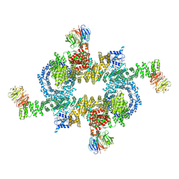
 | | Cryo-EM structure of the yeast spliceosome at 3.6 angstrom resolution | | 分子名称: | ADENOSINE-5'-DIPHOSPHATE, GUANOSINE-5'-DIPHOSPHATE, MAGNESIUM ION, ... | | 著者 | Yan, C, Hang, J, Wan, R, Huang, M, Wong, C, Shi, Y. | | 登録日 | 2015-08-09 | | 公開日 | 2015-09-23 | | 最終更新日 | 2024-03-20 | | 実験手法 | ELECTRON MICROSCOPY (3.6 Å) | | 主引用文献 | Structure of a yeast spliceosome at 3.6-angstrom resolution
Science, 349, 2015
|
|
3JCM
 | | Cryo-EM structure of the spliceosomal U4/U6.U5 tri-snRNP | | 分子名称: | 13 kDa ribonucleoprotein-associated protein, GUANOSINE-5'-TRIPHOSPHATE, N,N,7-trimethylguanosine 5'-(trihydrogen diphosphate), ... | | 著者 | Wan, R, Yan, C, Bai, R, Wang, L, Huang, M, Wong, C.C, Shi, Y. | | 登録日 | 2015-12-23 | | 公開日 | 2016-02-24 | | 最終更新日 | 2024-03-20 | | 実験手法 | ELECTRON MICROSCOPY (3.8 Å) | | 主引用文献 | The 3.8 angstrom structure of the U4/U6.U5 tri-snRNP: Insights into spliceosome assembly and catalysis
Science, 351, 2016
|
|
3MHW
 | | The complex crystal Structure of Urokianse and 2-Aminobenzothiazole | | 分子名称: | 1,3-benzothiazol-2-amine, SULFATE ION, Urokinase-type plasminogen activator | | 著者 | Jiang, L.-G, Yuan, C, Chen, L.-Q, Huang, M.-D. | | 登録日 | 2010-04-09 | | 公開日 | 2010-04-21 | | 最終更新日 | 2023-11-01 | | 実験手法 | X-RAY DIFFRACTION (1.45 Å) | | 主引用文献 | Crystal Structures of 2-Aminobenzothiazole-based Inhibitors in Complexes with Urokinase-type Plasminogen Activator
CHIN.J.STRUCT.CHEM., 28, 2009
|
|
5H64
 | | Cryo-EM structure of mTORC1 | | 分子名称: | Regulatory-associated protein of mTOR, Serine/threonine-protein kinase mTOR, Target of rapamycin complex subunit LST8 | | 著者 | Yang, H, Wang, J, Liu, M, Chen, X, Huang, M, Tan, D, Dong, M, Wong, C.C.L, Wang, J, Xu, Y, Wang, H. | | 登録日 | 2016-11-10 | | 公開日 | 2017-01-25 | | 最終更新日 | 2019-11-06 | | 実験手法 | ELECTRON MICROSCOPY (4.4 Å) | | 主引用文献 | 4.4 angstrom Resolution Cryo-EM structure of human mTOR Complex 1
Protein Cell, 7, 2016
|
|
6LBA
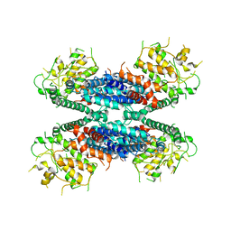 | | Cryo-EM structure of the AtMLKL2 tetramer | | 分子名称: | Protein kinase family protein | | 著者 | Lisa, M, Huang, M, Zhang, X, Ryohei, T.N, Leila, B.K, Isabel, M.L.S, Florence, J, Viera, K, Dmitry, L, Jane, E.P, James, M.M, Kay, H, Paul, S.L, Chai, J, Takaki, M. | | 登録日 | 2019-11-13 | | 公開日 | 2020-11-18 | | 最終更新日 | 2024-03-27 | | 実験手法 | ELECTRON MICROSCOPY (4.1 Å) | | 主引用文献 | Cryo-EM structure of the AtMLKL3 tetramer
To Be Published
|
|
1I9E
 | | TCR DOMAIN | | 分子名称: | 2-acetamido-2-deoxy-beta-D-glucopyranose, CYTOTOXIC TCELL VALPHA DOMAIN | | 著者 | Rudolph, M.G, Huang, M, Teyton, L, Wilson, I.A. | | 登録日 | 2001-03-19 | | 公開日 | 2001-12-05 | | 最終更新日 | 2023-08-09 | | 実験手法 | X-RAY DIFFRACTION (2.5 Å) | | 主引用文献 | Crystal structure of an isolated V(alpha) domain of the 2C T-cell receptor.
J.Mol.Biol., 314, 2001
|
|
1LEG
 | | Crystal Structure of H-2Kb bound to the dEV8 peptide | | 分子名称: | 2-acetamido-2-deoxy-beta-D-glucopyranose, 2-acetamido-2-deoxy-beta-D-glucopyranose-(1-4)-[alpha-L-fucopyranose-(1-6)]2-acetamido-2-deoxy-beta-D-glucopyranose, BETA-2-MICROGLOBULIN, ... | | 著者 | Luz, J.G, Huang, M, Garcia, K.C, Rudolph, M.G, Apostolopoulos, V, Teyton, L, Wilson, I.A. | | 登録日 | 2002-04-09 | | 公開日 | 2002-06-19 | | 最終更新日 | 2020-07-29 | | 実験手法 | X-RAY DIFFRACTION (1.75 Å) | | 主引用文献 | Structural comparison of allogeneic and syngeneic T cell receptor-peptide-major histocompatibility complex complexes: a buried alloreactive mutation subtly alters peptide presentation substantially increasing V(beta) Interactions.
J.Exp.Med., 195, 2002
|
|
3CDZ
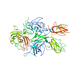 | | Crystal structure of human factor VIII | | 分子名称: | 2-acetamido-2-deoxy-beta-D-glucopyranose, 2-acetamido-2-deoxy-beta-D-glucopyranose-(1-4)-2-acetamido-2-deoxy-beta-D-glucopyranose, CALCIUM ION, ... | | 著者 | Ngo, J.C, Huang, M, Roth, D.A, Furie, B.C, Furie, B. | | 登録日 | 2008-02-27 | | 公開日 | 2008-04-01 | | 最終更新日 | 2023-08-30 | | 実験手法 | X-RAY DIFFRACTION (3.98 Å) | | 主引用文献 | Crystal structure of human factor VIII: implications for the formation of the factor IXa-factor VIIIa complex.
Structure, 16, 2008
|
|
3CX9
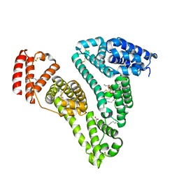 | | Crystal Structure of Human serum albumin complexed with Myristic acid and lysophosphatidylethanolamine | | 分子名称: | (2S)-3-{[(R)-(2-aminoethoxy)(hydroxy)phosphoryl]oxy}-2-hydroxypropyl hexadecanoate, MYRISTIC ACID, Serum albumin | | 著者 | Guo, S, Yang, F, Chen, L, Bian, C, Huang, M. | | 登録日 | 2008-04-24 | | 公開日 | 2009-04-28 | | 最終更新日 | 2023-11-01 | | 実験手法 | X-RAY DIFFRACTION (2.8 Å) | | 主引用文献 | Structural basis of transport of lysophospholipids by human serum albumin.
Biochem.J., 423, 2009
|
|
5H7V
 | |
1LEK
 | | Crystal Structure of H-2Kbm3 bound to dEV8 | | 分子名称: | 2-acetamido-2-deoxy-beta-D-glucopyranose, 2-acetamido-2-deoxy-beta-D-glucopyranose-(1-4)-[alpha-L-fucopyranose-(1-6)]2-acetamido-2-deoxy-beta-D-glucopyranose, Beta-2-microglobulin, ... | | 著者 | Luz, J.G, Huang, M, Garcia, K.C, Rudolph, M.G, Apostolopoulos, V, Teyton, L, Wilson, I.A. | | 登録日 | 2002-04-09 | | 公開日 | 2002-06-26 | | 最終更新日 | 2020-07-29 | | 実験手法 | X-RAY DIFFRACTION (2.15 Å) | | 主引用文献 | Structural comparison of allogeneic and syngeneic T cell receptor-peptide-major histocompatibility complex complexes: a buried alloreactive mutation subtly alters peptide presentation substantially increasing V(beta) Interactions.
J.Exp.Med., 195, 2002
|
|
6L05
 | | Crystal structure of uPA_H99Y in complex with 50F | | 分子名称: | 3-azanyl-5-(azepan-1-yl)-6-(1-benzofuran-2-yl)-Ncarbamimidoyl-pyrazine-2-carboxamide, Urokinase-type plasminogen activator | | 著者 | Buckley, B, Jiang, L.G, Huang, M.D, Kelso, M, Ranson, M. | | 登録日 | 2019-09-26 | | 公開日 | 2020-09-30 | | 最終更新日 | 2023-11-22 | | 実験手法 | X-RAY DIFFRACTION (2.49 Å) | | 主引用文献 | Crystal structure of uPA_H99Y in complex with 50F
To Be Published
|
|
6L04
 | | Crystal structure of uPA_H99Y in complex with 31F | | 分子名称: | 3-azanyl-5-(azepan-1-yl)-N-carbamimidoyl-6-(2,4-dimethoxypyrimidin-5-yl)pyrazine-2-carboxamide, Urokinase-type plasminogen activator | | 著者 | Buckley, B, Jiang, L.G, Huang, M.D, Kelso, M, Ranson, M. | | 登録日 | 2019-09-26 | | 公開日 | 2020-09-30 | | 最終更新日 | 2023-11-22 | | 実験手法 | X-RAY DIFFRACTION (2.21 Å) | | 主引用文献 | Crystal structure of uPA_H99Y in complex with 31F
To Be Published
|
|
4QTH
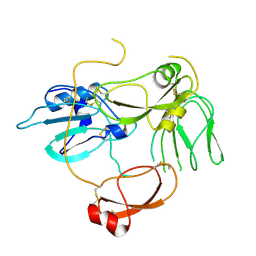
 | | Crystal structure of anti-uPAR Fab 8B12 | | 分子名称: | anti-uPAR antibody, heavy chain, light chain | | 著者 | Zhao, B, Yuan, C, Luo, Z, Huang, M. | | 登録日 | 2014-07-08 | | 公開日 | 2015-02-25 | | 最終更新日 | 2022-08-24 | | 実験手法 | X-RAY DIFFRACTION (2.17 Å) | | 主引用文献 | Stabilizing a flexible interdomain hinge region harboring the SMB binding site drives uPAR into its closed conformation.
J.Mol.Biol., 427, 2015
|
|


