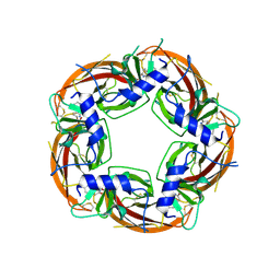
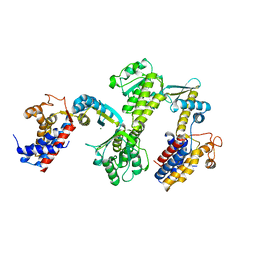
2XW6
 
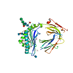
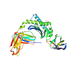
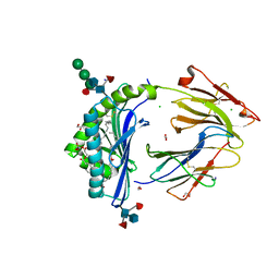
 | | The Crystal Structure of Methylglyoxal Synthase from Thermus sp. GH5 Bound to Phosphate Ion. | | 分子名称: | METHYLGLYOXAL SYNTHASE, PHOSPHATE ION | | 著者 | Shahsavar, A, Erfani Moghaddam, M, Antonyuk, S.V, Khajeh, K, Naderi-Manesh, H. | | 登録日 | 2010-11-01 | | 公開日 | 2011-11-09 | | 最終更新日 | 2023-12-20 | | 実験手法 | X-RAY DIFFRACTION (1.08 Å) | | 主引用文献 | Atomic Resolution Structure of Methylglyoxal Synthase from Thermus Sp. Gh5 Bound to Phosphate: Insights Into the Distinctive Effects of Phosphate on the Enzyme Structure
To be Published
|
|
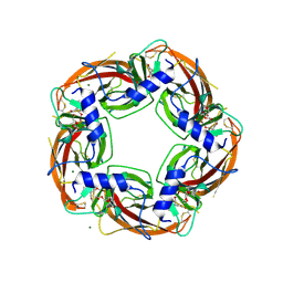
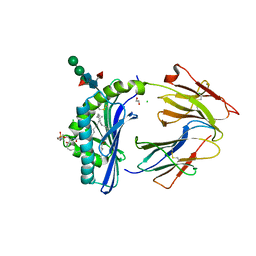
6ZBV
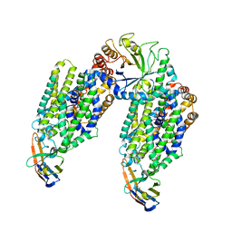
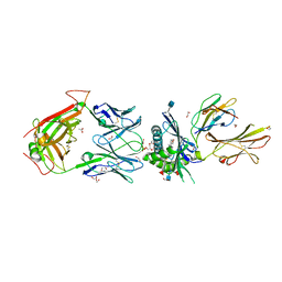
 | | Inward-open structure of human glycine transporter 1 in complex with a benzoylisoindoline inhibitor and sybody Sb_GlyT1#7 | | 分子名称: | Sodium- and chloride-dependent glycine transporter 1,Sodium- and chloride-dependent glycine transporter 1, Sybody Sb_GlyT1#7, [5-fluoranyl-6-(oxan-4-yloxy)-1,3-dihydroisoindol-2-yl]-[5-methylsulfonyl-2-[2,2,3,3,3-pentakis(fluoranyl)propoxy]phenyl]methanone | | 著者 | Shahsavar, A, Stohler, P, Bourenkov, G, Zimmermann, I, Siegrist, M, Guba, W, Pinard, E, Sinning, S, Seeger, M.A, Schneider, T.R, Dawson, R.J.P, Nissen, P. | | 登録日 | 2020-06-09 | | 公開日 | 2021-03-17 | | 最終更新日 | 2024-01-24 | | 実験手法 | X-RAY DIFFRACTION (3.4 Å) | | 主引用文献 | Structural insights into the inhibition of glycine reuptake.
Nature, 591, 2021
|
|
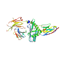
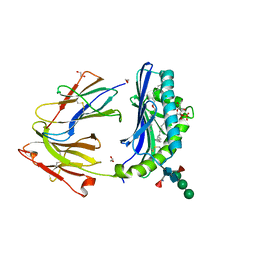
4UM3
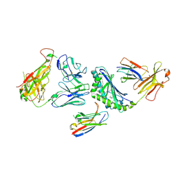
 | | Engineered Ls-AChBP with alpha4-alpha4 binding pocket in complex with NS3920 | | 分子名称: | 1-(6-bromopyridin-3-yl)-1,4-diazepane, 2-acetamido-2-deoxy-beta-D-glucopyranose, ACETYLCHOLINE BINDING PROTEIN, ... | | 著者 | Shahsavar, A, Kastrup, J.S, Balle, T, Gajhede, M. | | 登録日 | 2014-05-14 | | 公開日 | 2015-07-22 | | 最終更新日 | 2024-01-10 | | 実験手法 | X-RAY DIFFRACTION (2.703 Å) | | 主引用文献 | Achbp Engineered to Mimic the Alpha4-Alpha4 Binding Pocket in Alpha4Beta2 Nicotinic Acetylcholine Receptors Reveals Interface Specific Interactions Important for Binding and Activity
Mol.Pharmacol., 88, 2015
|
|
4UM1
 | | Engineered Ls-AChBP with alpha4-alpha4 binding pocket in complex with NS3573 | | 分子名称: | 1-(5-ethoxypyridin-3-yl)-1,4-diazepane, 2-acetamido-2-deoxy-beta-D-glucopyranose, ACETYLCHOLINE-BINDING PROTEIN | | 著者 | Shahsavar, A, Kastrup, J.S, Balle, T, Gajhede, M. | | 登録日 | 2014-05-14 | | 公開日 | 2015-07-22 | | 最終更新日 | 2024-01-10 | | 実験手法 | X-RAY DIFFRACTION (2.83 Å) | | 主引用文献 | Achbp Engineered to Mimic the Alpha4-Alpha4 Binding Pocket in Alpha4Beta2 Nicotinic Acetylcholine Receptors Reveals Interface Specific Interactions Important for Binding and Activity
Mol.Pharmacol., 88, 2015
|
|
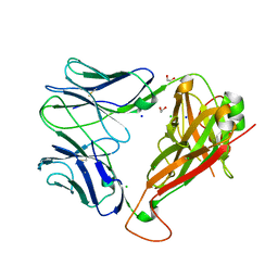
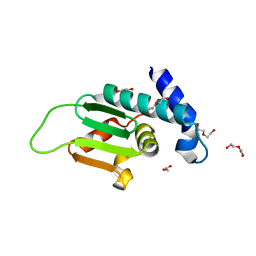
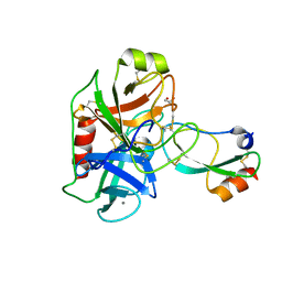
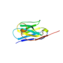
4OTK
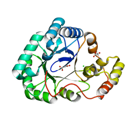 | | A structural characterization of the isoniazid Mycobacterium tuberculosis drug target, Rv2971, in its unliganded form | | 分子名称: | CHLORIDE ION, MALONATE ION, Mycobacterial Enzyme Rv2971 | | 著者 | Shahine, A, Beddoe, T. | | 登録日 | 2014-02-13 | | 公開日 | 2014-05-07 | | 最終更新日 | 2023-11-08 | | 実験手法 | X-RAY DIFFRACTION (1.6 Å) | | 主引用文献 | A structural characterization of the isoniazid Mycobacterium tuberculosis drug target, Rv2971, in its unliganded form
Acta Crystallogr.,Sect.F, 70, 2014
|
|
8GK6
 | |
6ZPL
 | | Inward-open structure of human glycine transporter 1 in complex with a benzoylisoindoline inhibitor, sybody Sb_GlyT1#7 and bound Na and Cl ions. | | 分子名称: | CHLORIDE ION, Endoglucanase H, SODIUM ION, ... | | 著者 | Shahsavar, A, Stohler, P, Bourenkov, G, Zimmermann, I, Siegrist, M, Guba, W, Pinard, E, Sinning, S, Seeger, M.A, Schneider, T.R, Dawson, R.J.P, Nissen, P. | | 登録日 | 2020-07-08 | | 公開日 | 2021-03-17 | | 最終更新日 | 2024-01-31 | | 実験手法 | X-RAY DIFFRACTION (3.945 Å) | | 主引用文献 | Structural insights into the inhibition of glycine reuptake.
Nature, 591, 2021
|
|
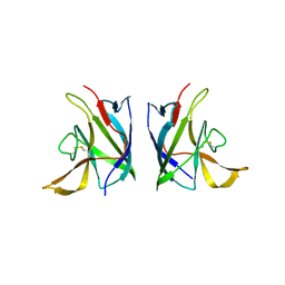
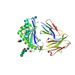
5WJO
 | | Crystal structure of the unliganded PG90 TCR | | 分子名称: | 1,2-ETHANEDIOL, CHLORIDE ION, PG90 TCR alpha chain, ... | | 著者 | Shahine, A, Gras, S, Rossjohn, J. | | 登録日 | 2017-07-24 | | 公開日 | 2017-10-25 | | 最終更新日 | 2023-10-04 | | 実験手法 | X-RAY DIFFRACTION (2.5 Å) | | 主引用文献 | A molecular basis of human T cell receptor autoreactivity toward self-phospholipids.
Sci Immunol, 2, 2017
|
|
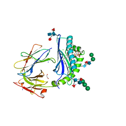
5WKG
 | | Crystal Structure of Human CD1b in Complex with PA | | 分子名称: | (2R)-1-(hexadecanoyloxy)-3-(phosphonooxy)propan-2-yl (9Z)-octadec-9-enoate, 1,2-ETHANEDIOL, 2-acetamido-2-deoxy-beta-D-glucopyranose, ... | | 著者 | Shahine, A, Gras, S, Rossjohn, J. | | 登録日 | 2017-07-25 | | 公開日 | 2017-11-01 | | 最終更新日 | 2020-07-29 | | 実験手法 | X-RAY DIFFRACTION (2.06 Å) | | 主引用文献 | A molecular basis of human T cell receptor autoreactivity toward self-phospholipids.
Sci Immunol, 2, 2017
|
|
5WL1
 | | Crystal Structure of Human CD1b in Complex with PG | | 分子名称: | (19S,22R,25R)-22,25,26-trihydroxy-16,22-dioxo-17,21,23-trioxa-22lambda~5~-phosphahexacosan-19-yl (9E)-octadec-9-enoate, 1,2-ETHANEDIOL, Beta-2-microglobulin, ... | | 著者 | Shahine, A, Gras, S, Rossjohn, J. | | 登録日 | 2017-07-25 | | 公開日 | 2017-11-01 | | 最終更新日 | 2020-07-29 | | 実験手法 | X-RAY DIFFRACTION (1.38 Å) | | 主引用文献 | A molecular basis of human T cell receptor autoreactivity toward self-phospholipids.
Sci Immunol, 2, 2017
|
|
5WKE
 | | Crystal Structure of Human CD1b in Complex with PS | | 分子名称: | (19S,22S,25R)-25-amino-22-hydroxy-16,22,26-trioxo-17,21,23-trioxa-22lambda~5~-phosphahexacosan-19-yl (9Z)-octadec-9-enoate, 1,2-ETHANEDIOL, 2-acetamido-2-deoxy-beta-D-glucopyranose, ... | | 著者 | Shahine, A, Gras, S, Rossjohn, J. | | 登録日 | 2017-07-25 | | 公開日 | 2017-11-01 | | 最終更新日 | 2020-07-29 | | 実験手法 | X-RAY DIFFRACTION (1.69 Å) | | 主引用文献 | A molecular basis of human T cell receptor autoreactivity toward self-phospholipids.
Sci Immunol, 2, 2017
|
|
5WKI
 | | Crystal structure of PG90 TCR-CD1b-PG complex | | 分子名称: | (19S,22R,25R)-22,25,26-trihydroxy-16,22-dioxo-17,21,23-trioxa-22lambda~5~-phosphahexacosan-19-yl (9E)-octadec-9-enoate, 1,2-ETHANEDIOL, 2-acetamido-2-deoxy-beta-D-glucopyranose, ... | | 著者 | Shahine, A, Gras, S, Rossjohn, J. | | 登録日 | 2017-07-25 | | 公開日 | 2017-11-01 | | 最終更新日 | 2020-07-29 | | 実験手法 | X-RAY DIFFRACTION (2.75 Å) | | 主引用文献 | A molecular basis of human T cell receptor autoreactivity toward self-phospholipids.
Sci Immunol, 2, 2017
|
|
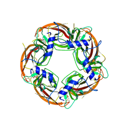
2X8W
 | | The Crystal Structure of Methylglyoxal Synthase from Thermus sp. GH5 Bound to Malonate. | | 分子名称: | MALONATE ION, METHYLGLYOXAL SYNTHASE | | 著者 | Shahsavar, A, Erfani Moghaddam, M, Antonyuk, S.V, Khajeh, K, Naderi-Manesh, H. | | 登録日 | 2010-03-13 | | 公開日 | 2011-03-23 | | 最終更新日 | 2023-12-20 | | 実験手法 | X-RAY DIFFRACTION (1.95 Å) | | 主引用文献 | Crystal Structures of Methylglyoxal Synthase from Thermus Sp.Gh5 in the Open and Closed Conformational States Provide Insight Into the Mechanism of Allosteric Regulation
To be Published
|
|
6V80
 | |
6V7Z
 | |
4ALX
 | | Crystal Structure of Ls-AChBP complexed with the potent nAChR antagonist DHbE | | 分子名称: | (4bS,6S)-6-methoxy-1,4,6,7,9,10,12,13-octahydro-3H,5H-pyrano[4',3':3,4]pyrido[2,1-i]indol-3-one, ACETYLCHOLINE BINDING PROTEIN, MAGNESIUM ION, ... | | 著者 | Shahsavar, A, Kastrup, J.S, Nielsen, E.O, Kristensen, J.L, Gajhede, M, Balle, T. | | 登録日 | 2012-03-06 | | 公開日 | 2012-08-29 | | 最終更新日 | 2018-01-17 | | 実験手法 | X-RAY DIFFRACTION (2.3 Å) | | 主引用文献 | Crystal Structure of Lymnaea Stagnalis Achbp Complexed with the Potent Nachr Antagonist Dh-Betab-E Suggests a Unique Mode of Antagonism
Plos One, 7, 2012
|
|
6V7Y
 | |
4ZY7
 | |
3RTK
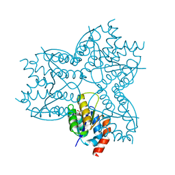
 | | Crystal structure of Cpn60.2 from Mycobacterium tuberculosis at 2.8A | | 分子名称: | 60 kDa chaperonin 2, MAGNESIUM ION | | 著者 | Shahar, A, Melamed-Frank, M, Kashi, Y, Adir, N. | | 登録日 | 2011-05-03 | | 公開日 | 2011-08-10 | | 最終更新日 | 2023-09-13 | | 実験手法 | X-RAY DIFFRACTION (2.8 Å) | | 主引用文献 | The dimeric structure of the Cpn60.2 chaperonin of Mycobacterium tuberculosis at 2.8 A reveals possible modes of function.
J.Mol.Biol., 412, 2011
|
|
8GLF
 | |
8GLE
 | | Crystal Structure of Human CD1b in Complex with Lysosulfatide | | 分子名称: | (2S,3R,4E)-2-amino-3-hydroxyoctadec-4-en-1-yl 3-O-sulfo-beta-D-galactopyranoside, 1,2-ETHANEDIOL, 2-acetamido-2-deoxy-beta-D-glucopyranose-(1-4)-[alpha-L-fucopyranose-(1-6)]2-acetamido-2-deoxy-beta-D-glucopyranose, ... | | 著者 | Shahine, A. | | 登録日 | 2023-03-22 | | 公開日 | 2023-09-20 | | 最終更新日 | 2024-05-01 | | 実験手法 | X-RAY DIFFRACTION (1.85 Å) | | 主引用文献 | CD1 lipidomes reveal lipid-binding motifs and size-based antigen-display mechanisms.
Cell, 186, 2023
|
|
6HAR
 | | Crystal structure of Mesotrypsin in complex with APPI-M17C/I18F/F34C | | 分子名称: | 1,2-ETHANEDIOL, Amyloid-beta A4 protein, CALCIUM ION, ... | | 著者 | Shahar, A, Cohen, I, Radisky, E, Papo, N. | | 登録日 | 2018-08-08 | | 公開日 | 2019-02-06 | | 最終更新日 | 2024-01-17 | | 実験手法 | X-RAY DIFFRACTION (1.497 Å) | | 主引用文献 | Disulfide engineering of human Kunitz-type serine protease inhibitors enhances proteolytic stability and target affinity toward mesotrypsin.
J.Biol.Chem., 294, 2019
|
|
8GLG
 | |
6XXO
 | |
6XXP
 | |


