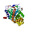
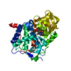
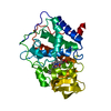
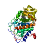
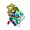
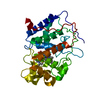
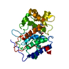
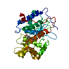
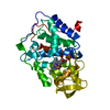
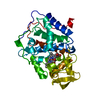
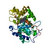
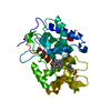
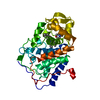
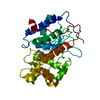
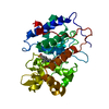
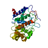
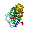
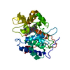
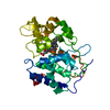
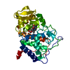
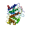
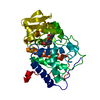
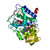
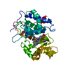
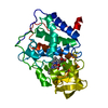
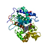
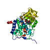
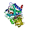
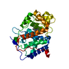
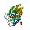
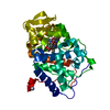
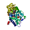
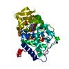
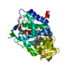
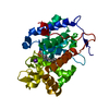
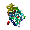
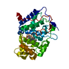
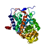
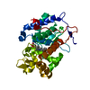
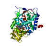
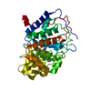
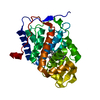
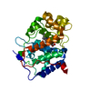
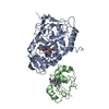
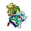
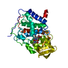
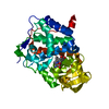
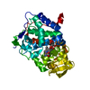
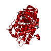
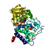
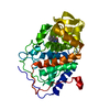
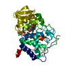
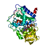
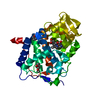
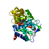
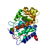
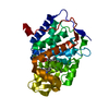
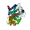
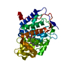
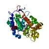
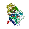
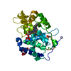
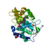
Entry Database : PDB / ID : 4a71Title cytochrome c peroxidase in complex with phenol CYTOCHROME C PEROXIDASE, MITOCHONDRIAL Keywords / / / / / / / / Function / homology Function Domain/homology Component
/ / / / / / / / / / / / / / / / / / / / / / / / / / / / / / / / / / Biological species SACCHAROMYCES CEREVISIAE (brewer's yeast)Method / / Resolution : 1.61 Å Authors Murphy, E.J. / Metcalfe, C.L. / Raven, E.L. / Moody, P.C.E. Journal : FEBS J. / Year : 2012Title : Crystal Structure of Guaiacol and Phenol Bound to a Heme Peroxidase.Authors : Murphy, E.J. / Metcalfe, C.L. / Nnamchi, C. / Moody, P.C.E. / Raven, E.L. History Deposition Nov 10, 2011 Deposition site / Processing site Revision 1.0 Oct 17, 2012 Provider / Type Revision 1.1 Jul 5, 2017 Group / Category / Item Revision 1.2 Dec 20, 2023 Group Data collection / Database references ... Data collection / Database references / Derived calculations / Other / Refinement description Category chem_comp_atom / chem_comp_bond ... chem_comp_atom / chem_comp_bond / database_2 / pdbx_database_status / pdbx_initial_refinement_model / struct_conn / struct_site Item _database_2.pdbx_DOI / _database_2.pdbx_database_accession ... _database_2.pdbx_DOI / _database_2.pdbx_database_accession / _pdbx_database_status.status_code_sf / _struct_conn.ptnr1_auth_comp_id / _struct_conn.ptnr1_auth_seq_id / _struct_conn.ptnr1_label_asym_id / _struct_conn.ptnr1_label_atom_id / _struct_conn.ptnr1_label_comp_id / _struct_conn.ptnr1_label_seq_id / _struct_conn.ptnr2_auth_comp_id / _struct_conn.ptnr2_auth_seq_id / _struct_conn.ptnr2_label_asym_id / _struct_conn.ptnr2_label_atom_id / _struct_conn.ptnr2_label_comp_id / _struct_conn.ptnr2_label_seq_id / _struct_site.pdbx_auth_asym_id / _struct_site.pdbx_auth_comp_id / _struct_site.pdbx_auth_seq_id
Show all Show less
 Movie
Movie Controller


 Open data
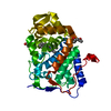
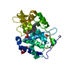
 Basic information
 Components
 Keywords
 Function and homology information
 X-RAY DIFFRACTION /
  Authors
 Citation
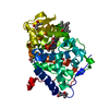
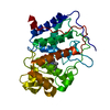
 Structure visualization
 Downloads & links
 Other downloads


 PDBj
PDBj

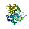
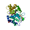
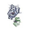
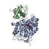
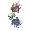
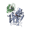
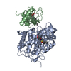
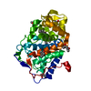
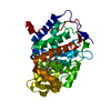
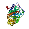
 Assembly
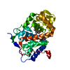


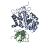
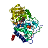
 Mass: 616.487 Da / Num. of mol.: 1 / Source method: obtained synthetically / Formula: C34H32FeN4O4
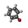
 Mass: 94.111 Da / Num. of mol.: 2 / Source method: obtained synthetically / Formula: C6H6O
 Mass: 18.015 Da / Num. of mol.: 494 / Source method: isolated from a natural source / Formula: H2O
 Sample preparation
 Processing