 Movie
Movie Controller

[English] 日本語
 Yorodumi
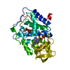
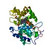
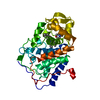
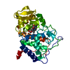
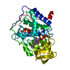
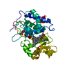
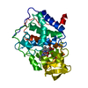
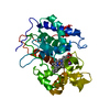
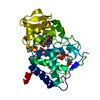
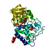
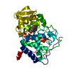
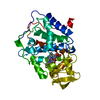
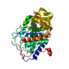
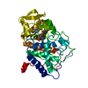
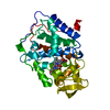
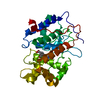
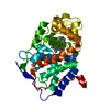
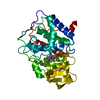
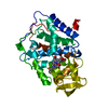
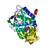
- PDB-1beq: INTERACTION BETWEEN PROXIMAL AND DISTALS REGIONS OF CYTOCHROME C ... -
+ Open data

- Basic information
| Entry | Database: PDB / ID: 1beq | ||||||
|---|---|---|---|---|---|---|---|
| Title | INTERACTION BETWEEN PROXIMAL AND DISTALS REGIONS OF CYTOCHROME C PEROXIDASE | ||||||
 Components Components | CYTOCHROME C PEROXIDASE | ||||||
 Keywords Keywords | PEROXIDASE / OXIDOREDUCTASE | ||||||
| Function / homology |  Function and homology information Function and homology informationcytochrome-c peroxidase / cytochrome-c peroxidase activity / hydrogen peroxide catabolic process / response to reactive oxygen species / peroxidase activity / mitochondrial intermembrane space / cellular response to oxidative stress / mitochondrial matrix / heme binding / mitochondrion / metal ion binding Similarity search - Function | ||||||
| Biological species |  | ||||||
| Method |  X-RAY DIFFRACTION / Resolution: 2.16 Å X-RAY DIFFRACTION / Resolution: 2.16 Å | ||||||
 Authors Authors | Miller, M.A. | ||||||
 Citation Citation | Journal: To be Published Title: Interaction between Proximal and Distals Regions of Cytochrome C Peroxidase Authors: Miller, M.A. / Han, G.W. / Kraut, J. #1: Journal: Nat.Struct.Biol. / Year: 1996Title: A Ligand-Gated, Hinged Loop Rearrangement Opens a Channel to a Buried Artificial Protein Cavity Authors: Fitzgerald, M.M. / Musah, R.A. / Mcree, D.E. / Goodin, D.B. #2: Journal: Proc.Natl.Acad.Sci.USA / Year: 1994Title: A Cation Binding Motif Stabilizes the Compound I Radical of Cytochrome C Peroxidase Authors: Miller, M.A. / Han, G.W. / Kraut, J. #3: Journal: Biochemistry / Year: 1990Title: X-Ray Structures of Recombinant Yeast Cytochrome C Peroxidase and Three Heme-Cleft Mutants Prepared by Site-Directed Mutagenesis Authors: Wang, J.M. / Mauro, M. / Edwards, S.L. / Oatley, S.J. / Fishel, L.A. / Ashford, V.A. / Xuong, N.H. / Kraut, J. #4: Journal: Biochemistry / Year: 1987Title: Yeast Cytochrome C Peroxidase: Mutagenesis and Expression in Escherichia Coli Show Tryptophan-51 is not the Radical Site in Compound I Authors: Fishel, L.A. / Villafranca, J.E. / Mauro, J.M. / Kraut, J. #5: Journal: J.Biol.Chem. / Year: 1984Title: Crystal Structure of Yeast Cytochrome C Peroxidase Refined at 1.7-A Resolution Authors: Finzel, B.C. / Poulos, T.L. / Kraut, J. | ||||||
| History |
|
- Structure visualization
| Structure viewer | Molecule: MolmilJmol/JSmol |
|---|
- Downloads & links
-Download
| PDBx/mmCIF format | 1beq.cif.gz | 78.9 KB | Display | PDBx/mmCIF format |
|---|---|---|---|---|
| PDB format | pdb1beq.ent.gz | 57.9 KB | Display | PDB format |
| PDBx/mmJSON format | 1beq.json.gz | Tree view | PDBx/mmJSON format | |
| Others |  Other downloads Other downloads |
-Validation report
| Arichive directory | https://data.pdbj.org/pub/pdb/validation_reports/be/1beqftp://data.pdbj.org/pub/pdb/validation_reports/be/1beq | HTTPS FTP |
|---|
-Related structure data


-Links
 PDBj
PDBj
- Assembly
| Deposited unit | 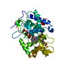
| ||||||||
|---|---|---|---|---|---|---|---|---|---|
| 1 |
| ||||||||
| Unit cell |
|
-Components
| #1: Protein | Mass: 33203.887 Da / Num. of mol.: 1 / Mutation: MET ILE ADDED AT N-TERMINUS, W191Y Source method: isolated from a genetically manipulated source Details: COMPLEXED WITH A MES (+) ZWITTERION Source: (gene. exp.) Production host:  |
|---|---|
| #2: Chemical | ChemComp-HEM /   Mass: 616.487 Da / Num. of mol.: 1 / Source method: obtained synthetically / Formula: C34H32FeN4O4 Mass: 616.487 Da / Num. of mol.: 1 / Source method: obtained synthetically / Formula: C34H32FeN4O4 |
| #3: Chemical | ChemComp-MES /   Mass: 195.237 Da / Num. of mol.: 1 / Source method: obtained synthetically / Formula: C6H13NO4S / Comment: pH buffer*YM Mass: 195.237 Da / Num. of mol.: 1 / Source method: obtained synthetically / Formula: C6H13NO4S / Comment: pH buffer*YM |
| #4: Water | ChemComp-HOH /  Mass: 18.015 Da / Num. of mol.: 173 / Source method: isolated from a natural source / Formula: H2O Mass: 18.015 Da / Num. of mol.: 173 / Source method: isolated from a natural source / Formula: H2O |
| Compound details | THE MUTATION DESTABILIZES THE 191 LOOP. IN MES BUFFER, A MOLECULE OF MES BINDS TO THE ENZYME, AND ...THE MUTATION DESTABILIZ |
| Sequence details | THIS CYTOCHROME C PEROXIDASE DIFFERS FROM A PREVIOUSLY DEPOSITED STRUCTURE (PROTEIN DATA BANK ENTRY ...THIS CYTOCHROME |
-Experimental details
-Experiment
| Experiment | Method: X-RAY DIFFRACTION / Number of used crystals: 1 |
|---|
- Sample preparation
| Crystal | Density Matthews: 2.63 Å3/Da / Density % sol: 53 % |
|---|---|
| Crystal grow | pH: 6 / Details: 30% MPD; 50 MM MES/TRIS, PH 6.0 |
-Data collection
| Diffraction | Mean temperature: 273 K |
|---|---|
| Diffraction source | Wavelength: 1.5418 |
| Detector | Date: Oct 1, 1997 |
| Radiation | Monochromatic (M) / Laue (L): M / Scattering type: x-ray |
| Radiation wavelength | Wavelength: 1.5418 Å / Relative weight: 1 |
| Reflection | Highest resolution: 2.16 Å |
- Processing
| Software | Name: TNT / Classification: refinement | ||||||||||||||||||||||||||||||
|---|---|---|---|---|---|---|---|---|---|---|---|---|---|---|---|---|---|---|---|---|---|---|---|---|---|---|---|---|---|---|---|
| Refinement | Resolution: 2.16→20 Å / Isotropic thermal model: BCORREL / σ(F): 2 / Stereochemistry target values: PROTGEO Details: COORDINATES FOR RESIDUES -1, 0, AND 1 - 3 ARE NOT INCLUDED IN THIS ENTRY BECAUSE THESE RESIDUES COULD NOT BE RESOLVED IN THE FINAL ELECTRON DENSITY MAPS. WATER MOLECULES WITH B-FACTORS ...Details: COORDINATES FOR RESIDUES -1, 0, AND 1 - 3 ARE NOT INCLUDED IN THIS ENTRY BECAUSE THESE RESIDUES COULD NOT BE RESOLVED IN THE FINAL ELECTRON DENSITY MAPS. WATER MOLECULES WITH B-FACTORS GREATER THAN 80 WERE NOT INCLUDED IN THE MODEL.
| ||||||||||||||||||||||||||||||
| Refinement step | Cycle: LAST / Resolution: 2.16→20 Å
| ||||||||||||||||||||||||||||||
| Refine LS restraints |
|
