 Movie
Movie Controller

[English] 日本語
 Yorodumi
- PDB-1bes: INTERACTION BETWEEN PROXIMAL AND DISTALS REGIONS OF CYTOCHROME C ... -
+ Open data

- Basic information
| Entry | Database: PDB / ID: 1bes | ||||||
|---|---|---|---|---|---|---|---|
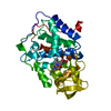
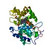
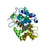
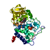
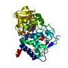
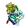
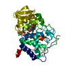
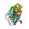
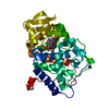
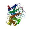
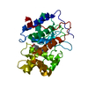
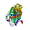
| Title | INTERACTION BETWEEN PROXIMAL AND DISTALS REGIONS OF CYTOCHROME C PEROXIDASE | ||||||
 Components Components | CYTOCHROME C PEROXIDASE | ||||||
 Keywords Keywords | PEROXIDASE / OXIDOREDUCTASE | ||||||
| Function / homology |  Function and homology information Function and homology informationcytochrome-c peroxidase / cytochrome-c peroxidase activity / hydrogen peroxide catabolic process / response to reactive oxygen species / peroxidase activity / mitochondrial intermembrane space / cellular response to oxidative stress / mitochondrial matrix / heme binding / mitochondrion / metal ion binding Similarity search - Function | ||||||
| Biological species |  | ||||||
| Method |  X-RAY DIFFRACTION / Resolution: 2 Å X-RAY DIFFRACTION / Resolution: 2 Å | ||||||
 Authors Authors | Miller, M.A. / Kraut, J. | ||||||
 Citation Citation | Journal: To be Published Title: Interaction between Proximal and Distals Regions of Cytochrome C Peroxidase Authors: Miller, M.A. / Han, G.W. / Kraut, J. #1: Journal: Nat.Struct.Biol. / Year: 1996Title: A Ligand-Gated, Hinged Loop Rearrangement Opens a Channel to a Buried Artificial Protein Cavity Authors: Fitzgerald, M.M. / Musah, R.A. / Mcree, D.E. / Goodin, D.B. #2: Journal: Proc.Natl.Acad.Sci.USA / Year: 1994Title: A Cation Binding Motif Stabilizes the Compound I Radical of Cytochrome C Peroxidase Authors: Miller, M.A. / Han, G.W. / Kraut, J. #3: Journal: Biochemistry / Year: 1990Title: X-Ray Structures of Recombinant Yeast Cytochrome C Peroxidase and Three Heme-Cleft Mutants Prepared by Site-Directed Mutagenesis Authors: Wang, J.M. / Mauro, M. / Edwards, S.L. / Oatley, S.J. / Fishel, L.A. / Ashford, V.A. / Xuong, N.H. / Kraut, J. #4: Journal: Biochemistry / Year: 1987Title: Yeast Cytochrome C Peroxidase: Mutagenesis and Expression in Escherichia Coli Show Tryptophan-51 is not the Radical Site in Compound I Authors: Fishel, L.A. / Villafranca, J.E. / Mauro, J.M. / Kraut, J. #5: Journal: J.Biol.Chem. / Year: 1984Title: Crystal Structure of Yeast Cytochrome C Peroxidase Refined at 1.7-A Resolution Authors: Finzel, B.C. / Poulos, T.L. / Kraut, J. | ||||||
| History |
|
- Structure visualization
| Structure viewer | Molecule: MolmilJmol/JSmol |
|---|
- Downloads & links
-Download
| PDBx/mmCIF format | 1bes.cif.gz | 79.6 KB | Display | PDBx/mmCIF format |
|---|---|---|---|---|
| PDB format | pdb1bes.ent.gz | 58.2 KB | Display | PDB format |
| PDBx/mmJSON format | 1bes.json.gz | Tree view | PDBx/mmJSON format | |
| Others |  Other downloads Other downloads |
-Validation report
| Arichive directory | https://data.pdbj.org/pub/pdb/validation_reports/be/1besftp://data.pdbj.org/pub/pdb/validation_reports/be/1bes | HTTPS FTP |
|---|
-Related structure data


-Links
 PDBj
PDBj
- Assembly
| Deposited unit | 
| ||||||||
|---|---|---|---|---|---|---|---|---|---|
| 1 |
| ||||||||
| Unit cell |
|
-Components
| #1: Protein | Mass: 33203.887 Da / Num. of mol.: 1 / Mutation: MET ILE ADDED AT N-TERMINUS, W191Y Source method: isolated from a genetically manipulated source Details: FE(III)-CL COMPLEX, WITH A MES (+) ZWITTERION BOUND IN THE PROXIMAL REGION Source: (gene. exp.) Plasmid: PUC8 / Production host:  |
|---|---|
| #2: Chemical | ChemComp-CL /   Mass: 35.453 Da / Num. of mol.: 1 / Source method: obtained synthetically / Formula: Cl Mass: 35.453 Da / Num. of mol.: 1 / Source method: obtained synthetically / Formula: Cl |
| #3: Chemical | ChemComp-HEM /   Mass: 616.487 Da / Num. of mol.: 1 / Source method: obtained synthetically / Formula: C34H32FeN4O4 Mass: 616.487 Da / Num. of mol.: 1 / Source method: obtained synthetically / Formula: C34H32FeN4O4 |
| #4: Chemical | ChemComp-MES /   Mass: 195.237 Da / Num. of mol.: 1 / Source method: obtained synthetically / Formula: C6H13NO4S / Comment: pH buffer*YM Mass: 195.237 Da / Num. of mol.: 1 / Source method: obtained synthetically / Formula: C6H13NO4S / Comment: pH buffer*YM |
| #5: Water | ChemComp-HOH /  Mass: 18.015 Da / Num. of mol.: 169 / Source method: isolated from a natural source / Formula: H2O Mass: 18.015 Da / Num. of mol.: 169 / Source method: isolated from a natural source / Formula: H2O |
| Compound details | THE MUTATION INFLUENCES THE COORDINATION STATE OF THE ENZYME. THE BINDING OF MES FROM BUFFER TENDS ...THE MUTATION INFLUENCES |
| Sequence details | THIS CYTOCHROME C PEROXIDASE DIFFERS FROM A PREVIOUSLY DEPOSITED STRUCTURE (PROTEIN DATA BANK ENTRY ...THIS CYTOCHROME |
-Experimental details
-Experiment
| Experiment | Method: X-RAY DIFFRACTION / Number of used crystals: 1 |
|---|
- Sample preparation
| Crystal | Density Matthews: 2.66 Å3/Da / Density % sol: 54 % |
|---|---|
| Crystal grow | pH: 6 / Details: 30% MPD, 20 MM TRIS/MES PH 6.0, 150 MM NACL |
-Data collection
| Diffraction | Mean temperature: 273 K |
|---|---|
| Diffraction source | Wavelength: 1.5418 |
| Detector | Date: Nov 1, 1997 |
| Radiation | Monochromatic (M) / Laue (L): M / Scattering type: x-ray |
| Radiation wavelength | Wavelength: 1.5418 Å / Relative weight: 1 |
| Reflection | Highest resolution: 2 Å / Num. obs: 21914 / % possible obs: 90 % / Rsym value: 0.065 |
- Processing
| Software | Name: TNT / Version: 1 / Classification: refinement | ||||||||||||||||||||||||||||||
|---|---|---|---|---|---|---|---|---|---|---|---|---|---|---|---|---|---|---|---|---|---|---|---|---|---|---|---|---|---|---|---|
| Refinement | Resolution: 2→20 Å / Isotropic thermal model: TNT BCORREL.DAT / σ(F): 2 / Stereochemistry target values: TNT PROTGEO Details: RESTRAINTS WERE RELAXED FOR PLANARITY OF THE BACKBONE UNITS FOR RESIDUES 191-195, OTHERWISE THE DENSITY COULD NOT BE MODELLED PROPERLY. THE PLANARITY OF THE PEPTIDE BOND WAS RELAXED FOR ...Details: RESTRAINTS WERE RELAXED FOR PLANARITY OF THE BACKBONE UNITS FOR RESIDUES 191-195, OTHERWISE THE DENSITY COULD NOT BE MODELLED PROPERLY. THE PLANARITY OF THE PEPTIDE BOND WAS RELAXED FOR RESIDUES 190-194 BECAUSE IT WAS THE ONLY WAY THE MODEL COULD BE FITTED INTO THE DENSITY. THE DENSITY WAS CONTINUOUS THROUGH THIS REGION, AND SO THE AUTHORS CHOSE TO MODEL INTO THE DENSITY RATHER THAN TO PRESERVE THE REQUIREMENT FOR PLANARITY OF OMEGA. COORDINATES FOR RESIDUES -1, 0, AND 1 - 3 ARE NOT INCLUDED IN THIS ENTRY BECAUSE THESE RESIDUES COULD NOT BE RESOLVED IN THE FINAL ELECTRON DENSITY MAPS. WATER MOLECULES WITH B-FACTORS GREATER THAN 80 WERE NOT INCLUDED IN THE MODEL.
| ||||||||||||||||||||||||||||||
| Refinement step | Cycle: LAST / Resolution: 2→20 Å
| ||||||||||||||||||||||||||||||
| Refine LS restraints |
|
