 Movie
Movie Controller

[English] 日本語
 Yorodumi
- PDB-1a2f: PROBING THE STRENGTH AND CHARACTER OF AN ASP-HIS-X HYDROGEN BOND ... -
+ Open data

- Basic information
| Entry | Database: PDB / ID: 1a2f | ||||||
|---|---|---|---|---|---|---|---|
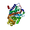
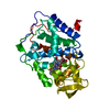
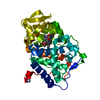
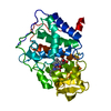
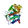
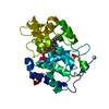
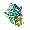
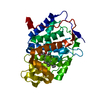
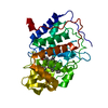
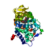
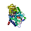
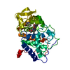
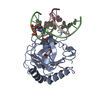
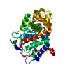
| Title | PROBING THE STRENGTH AND CHARACTER OF AN ASP-HIS-X HYDROGEN BOND BY INTRODUCING BURIED CHARGES | ||||||
 Components Components | CYTOCHROME C PEROXIDASE | ||||||
 Keywords Keywords | OXIDOREDUCTASE / PEROXIDASE | ||||||
| Function / homology |  Function and homology information Function and homology informationcytochrome-c peroxidase / cytochrome-c peroxidase activity / hydrogen peroxide catabolic process / response to reactive oxygen species / peroxidase activity / mitochondrial intermembrane space / cellular response to oxidative stress / mitochondrial matrix / heme binding / mitochondrion / metal ion binding Similarity search - Function | ||||||
| Biological species |  | ||||||
| Method |  X-RAY DIFFRACTION / MOLECULAR REPLACEMENT / Resolution: 2.1 Å X-RAY DIFFRACTION / MOLECULAR REPLACEMENT / Resolution: 2.1 Å | ||||||
 Authors Authors | Cao, Y. / Goodin, D.B. / Mcree, D.E. | ||||||
 Citation Citation | Journal: To be Published Title: Probing the Strength and Character of an Asp-His-X Hydrogen Bond by Introducing Buried Charges Authors: Cao, Y. / Musah, R.A. / Goodin, D.B. / Mcree, D.E. #1: Journal: Biochemistry / Year: 1993Title: The Asp-His-Fe Triad of Cytochrome C Peroxidase Controls the Reduction Potential, Electronic Structure, and Coupling of the Tryptophan Free Radical to the Heme Authors: Goodin, D.B. / Mcree, D.E. | ||||||
| History |
|
- Structure visualization
| Structure viewer | Molecule: MolmilJmol/JSmol |
|---|
- Downloads & links
-Download
| PDBx/mmCIF format | 1a2f.cif.gz | 86.5 KB | Display | PDBx/mmCIF format |
|---|---|---|---|---|
| PDB format | pdb1a2f.ent.gz | 64.9 KB | Display | PDB format |
| PDBx/mmJSON format | 1a2f.json.gz | Tree view | PDBx/mmJSON format | |
| Others |  Other downloads Other downloads |
-Validation report
| Arichive directory | https://data.pdbj.org/pub/pdb/validation_reports/a2/1a2fftp://data.pdbj.org/pub/pdb/validation_reports/a2/1a2f | HTTPS FTP |
|---|
-Related structure data
| Related structure data | 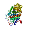 1a2gC 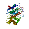 1cclC 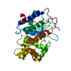 1ccaS S: Starting model for refinement C: citing same article ( |
|---|---|
| Similar structure data |


-Links
 PDBj
PDBj
- Assembly
| Deposited unit | 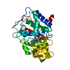
| ||||||||
|---|---|---|---|---|---|---|---|---|---|
| 1 |
| ||||||||
| Unit cell |
|
-Components
| #1: Protein | Mass: 33223.922 Da / Num. of mol.: 1 / Mutation: M172K Source method: isolated from a genetically manipulated source Source: (gene. exp.) Cell line: BL21 / Gene: CCP / Organelle: MITOCHONDRIA / Plasmid: PT7CCP / Species (production host): Escherichia coli / Cellular location (production host): CYTOPLASM / Gene (production host): CCP(MKT) / Production host:  |
|---|---|
| #2: Chemical | ChemComp-HEM /   Mass: 616.487 Da / Num. of mol.: 1 / Source method: obtained synthetically / Formula: C34H32FeN4O4 Mass: 616.487 Da / Num. of mol.: 1 / Source method: obtained synthetically / Formula: C34H32FeN4O4 |
| #3: Water | ChemComp-HOH /  Mass: 18.015 Da / Num. of mol.: 107 / Source method: isolated from a natural source / Formula: H2O Mass: 18.015 Da / Num. of mol.: 107 / Source method: isolated from a natural source / Formula: H2O |
-Experimental details
-Experiment
| Experiment | Method: X-RAY DIFFRACTION / Number of used crystals: 1 |
|---|
- Sample preparation
| Crystal | Density Matthews: 3.1 Å3/Da / Density % sol: 61 % |
|---|---|
| Crystal grow | Method: dialysis / Details: DIALYSIS AGAINST DISTILLED WATER, dialysis |
-Data collection
| Diffraction | Mean temperature: 290 K |
|---|---|
| Diffraction source | Source: ROTATING ANODE / Type: RIGAKU RUH2R / Wavelength: 1.5418 |
| Detector | Type: SIEMENS / Detector: AREA DETECTOR / Date: Jun 1, 1995 / Details: NO |
| Radiation | Monochromator: NO / Monochromatic (M) / Laue (L): M / Scattering type: x-ray |
| Radiation wavelength | Wavelength: 1.5418 Å / Relative weight: 1 |
| Reflection | Highest resolution: 2.1 Å / Num. obs: 24039 / % possible obs: 93.5 % / Observed criterion σ(I): 1.1 / Redundancy: 3.6 % / Biso Wilson estimate: 38.5 Å2 / Rmerge(I) obs: 0.09 / Rsym value: 0.089 / Net I/σ(I): 8.3 |
| Reflection shell | Resolution: 2.02→2.1 Å / Redundancy: 2.8 % / Mean I/σ(I) obs: 1 / Rsym value: 0.29 / % possible all: 81.6 |
- Processing
| Software |
| |||||||||||||||||||||
|---|---|---|---|---|---|---|---|---|---|---|---|---|---|---|---|---|---|---|---|---|---|---|
| Refinement | Method to determine structure: MOLECULAR REPLACEMENT Starting model: 1CCA Resolution: 2.1→5 Å / Data cutoff high absF: 100000 / Data cutoff low absF: 1 /
| |||||||||||||||||||||
| Refinement step | Cycle: LAST / Resolution: 2.1→5 Å
| |||||||||||||||||||||
| Xplor file |
|
