 Movie
Movie Controller

[English] 日本語
 Yorodumi
- PDB-2icv: Kinetic and Crystallographic Studies of a Redesigned Manganese-Bi... -
+ Open data

- Basic information
| Entry | Database: PDB / ID: 2icv | ||||||
|---|---|---|---|---|---|---|---|
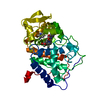
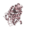
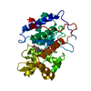
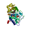
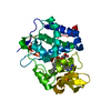
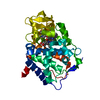
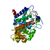
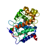
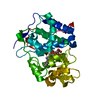
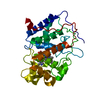
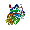
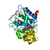
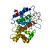
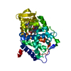
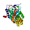
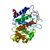
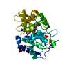
| Title | Kinetic and Crystallographic Studies of a Redesigned Manganese-Binding Site in Cytochrome c Peroxidase | ||||||
 Components Components | Cytochrome c peroxidase, mitochondrial | ||||||
 Keywords Keywords | OXIDOREDUCTASE / Manganese oxidation / metal-binding site / protein engineering / metalloprotein / biomimetic | ||||||
| Function / homology |  Function and homology information Function and homology informationcytochrome-c peroxidase / cytochrome-c peroxidase activity / response to reactive oxygen species / hydrogen peroxide catabolic process / peroxidase activity / mitochondrial intermembrane space / cellular response to oxidative stress / mitochondrial matrix / heme binding / mitochondrion / metal ion binding Similarity search - Function | ||||||
| Biological species |  | ||||||
| Method |  X-RAY DIFFRACTION / SYNCHROTRON / MOLECULAR REPLACEMENT / Resolution: 1.6 Å X-RAY DIFFRACTION / SYNCHROTRON / MOLECULAR REPLACEMENT / Resolution: 1.6 Å | ||||||
 Authors Authors | Pfister, T. / Mirarefi, A.Y. / Gengenbach, A.J. / Zhao, X. / Conaster, C.D.N. / Gao, Y.G. / Robinson, H. / Zukoski, C.F. / Wang, A.H.J. / Lu, Y. | ||||||
 Citation Citation | Journal: J.Biol.Inorg.Chem. / Year: 2007 Title: Kinetic and crystallographic studies of a redesigned manganese-binding site in cytochrome c peroxidase Authors: Pfister, T.D. / Mirarefi, A.Y. / Gengenbach, A.J. / Zhao, X. / Danstrom, C. / Conatser, N. / Gao, Y.-G. / Robinson, H. / Zukoski, C.F. / Wang, A.H.-J. / Lu, Y. | ||||||
| History |
|
- Structure visualization
| Structure viewer | Molecule: MolmilJmol/JSmol |
|---|
- Downloads & links
-Download
| PDBx/mmCIF format | 2icv.cif.gz | 75.5 KB | Display | PDBx/mmCIF format |
|---|---|---|---|---|
| PDB format | pdb2icv.ent.gz | 54.7 KB | Display | PDB format |
| PDBx/mmJSON format | 2icv.json.gz | Tree view | PDBx/mmJSON format | |
| Others |  Other downloads Other downloads |
-Validation report
| Arichive directory | https://data.pdbj.org/pub/pdb/validation_reports/ic/2icvftp://data.pdbj.org/pub/pdb/validation_reports/ic/2icv | HTTPS FTP |
|---|
-Related structure data
| Related structure data | 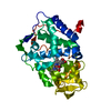 2ia8C 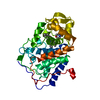 1besS S: Starting model for refinement C: citing same article ( |
|---|---|
| Similar structure data |


-Links
 PDBj
PDBj

- Assembly
| Deposited unit | 
| ||||||||
|---|---|---|---|---|---|---|---|---|---|
| 1 |
| ||||||||
| Unit cell |
|
-Components
| #1: Protein | Mass: 33260.910 Da / Num. of mol.: 1 Source method: isolated from a genetically manipulated source Source: (gene. exp.) Gene: CCP1 / Production host:  |
|---|---|
| #2: Chemical | ChemComp-CO /   Mass: 58.933 Da / Num. of mol.: 1 / Source method: obtained synthetically / Formula: Co Mass: 58.933 Da / Num. of mol.: 1 / Source method: obtained synthetically / Formula: Co |
| #3: Chemical | ChemComp-HEM /   Mass: 616.487 Da / Num. of mol.: 1 / Source method: obtained synthetically / Formula: C34H32FeN4O4 Mass: 616.487 Da / Num. of mol.: 1 / Source method: obtained synthetically / Formula: C34H32FeN4O4 |
| #4: Water | ChemComp-HOH /  Mass: 18.015 Da / Num. of mol.: 97 / Source method: isolated from a natural source / Formula: H2O Mass: 18.015 Da / Num. of mol.: 97 / Source method: isolated from a natural source / Formula: H2O |
-Experimental details
-Experiment
| Experiment | Method: X-RAY DIFFRACTION / Number of used crystals: 1 |
|---|
- Sample preparation
| Crystal | Density Matthews: 2.36 Å3/Da / Density % sol: 47.91 % |
|---|---|
| Crystal grow | Temperature: 277 K / Method: vapor diffusion, hanging drop / pH: 4.6 Details: 1.0 M hexanediol in 0.1 M sodium acetate trihydrate, VAPOR DIFFUSION, HANGING DROP, temperature 277K, pH 4.6 |
-Data collection
| Diffraction | Mean temperature: 95 K |
|---|---|
| Diffraction source | Source: SYNCHROTRON / Site: NSLS  / Beamline: X12C / Wavelength: 1.1 Å / Beamline: X12C / Wavelength: 1.1 Å |
| Detector | Type: ADSC QUANTUM 210 / Detector: CCD / Date: Jan 1, 2003 |
| Radiation | Monochromator: graphite / Protocol: SINGLE WAVELENGTH / Monochromatic (M) / Laue (L): M / Scattering type: x-ray |
| Radiation wavelength | Wavelength: 1.1 Å / Relative weight: 1 |
| Reflection | Resolution: 1.6→30 Å / Num. all: 35391 / Num. obs: 34426 / % possible obs: 83.1 % / Observed criterion σ(F): 0 / Observed criterion σ(I): 0 / Redundancy: 6.1 % / Rmerge(I) obs: 0.048 |
| Reflection shell | Resolution: 1.6→30 Å / Redundancy: 4.9 % / Rmerge(I) obs: 0.302 / % possible all: 83.1 |
- Processing
| Software |
| ||||||||||||||||||
|---|---|---|---|---|---|---|---|---|---|---|---|---|---|---|---|---|---|---|---|
| Refinement | Method to determine structure: MOLECULAR REPLACEMENT Starting model: PDB Entry: 1BES Resolution: 1.6→10 Å / σ(F): 4 / σ(I): 2
| ||||||||||||||||||
| Refinement step | Cycle: LAST / Resolution: 1.6→10 Å
| ||||||||||||||||||
| Refine LS restraints |
|
