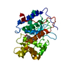
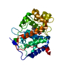
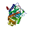
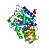
- PDB-4jmv: Crystal structure of Cytochrome C Peroxidase W191G-Gateless in co... -
+
Open data
ID or keywords:
Loading...
-
Basic information
Entry
Database: PDB / ID: 4jmv
Title
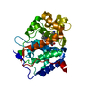
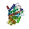
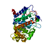
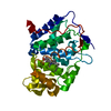
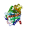
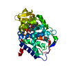
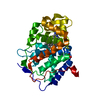
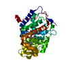
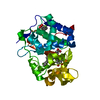
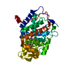
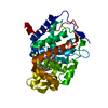
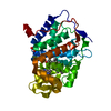
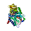
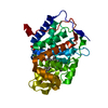
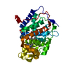
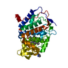
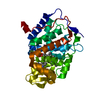
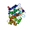
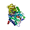
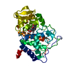
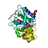
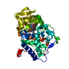
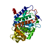
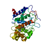
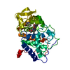
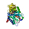
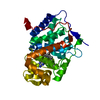
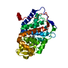
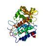
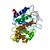
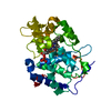
Crystal structure of Cytochrome C Peroxidase W191G-Gateless in complex with imidazo[1,2-a]pyridin-6-amine
Components
Cytochrome c peroxidase
Keywords
OXIDOREDUCTASE / Model system / bulk solvent / ordered waters / docking / ligand binding
Function / homology
Function and homology information
cytochrome-c peroxidase activity / Oxidoreductases; Acting on a peroxide as acceptor; Peroxidases / response to reactive oxygen species / hydrogen peroxide catabolic process / mitochondrial intermembrane space / cellular response to oxidative stress / mitochondrial matrix / heme binding / metal ion binding Similarity search - Function
Class I peroxidase / Heme-binding peroxidase Ccp1-like / Peroxidase; domain 2 / Peroxidase, domain 2 / Peroxidase; domain 1 - #10 / Peroxidase; domain 1 / Peroxidases heam-ligand binding site / Peroxidase, active site / Peroxidases active site signature. / Plant heme peroxidase family profile. ...Class I peroxidase / Heme-binding peroxidase Ccp1-like / Peroxidase; domain 2 / Peroxidase, domain 2 / Peroxidase; domain 1 - #10 / Peroxidase; domain 1 / Peroxidases heam-ligand binding site / Peroxidase, active site / Peroxidases active site signature. / Plant heme peroxidase family profile. / Haem peroxidase / Peroxidase / Peroxidases proximal heme-ligand signature. / Haem peroxidase superfamily / Orthogonal Bundle / Mainly Alpha Similarity search - Domain/homology
Mass: 18.015 Da / Num. of mol.: 414 / Source method: isolated from a natural source / Formula: H2O
-
Experimental details
-
Experiment
Experiment
Method: X-RAY DIFFRACTION / Number of used crystals: 1
-
Sample preparation
Crystal
Density Matthews: 3.06 Å3/Da / Density % sol: 59.81 %
Crystal grow
Temperature: 283 K / Method: vapor diffusion, sitting drop / pH: 6 Details: Apo crystal grown in 100mM KPi, pH 6.0. Soaked into 100mM ZINC18555973 in 25% MPD (30min), vapor diffusion, sitting drop, temperature 283K, VAPOR DIFFUSION, SITTING DROP
-
Data collection
Diffraction
Mean temperature: 80 K
Diffraction source
Source: SYNCHROTRON / Site: ALS / Beamline: 8.3.1 / Wavelength: 1.115869 Å
In the structure databanks used in Yorodumi, some data are registered as the other names, "COVID-19 virus" and "2019-nCoV". Here are the details of the virus and the list of structure data.
Jan 31, 2019. EMDB accession codes are about to change! (news from PDBe EMDB page)
EMDB accession codes are about to change! (news from PDBe EMDB page)
The allocation of 4 digits for EMDB accession codes will soon come to an end. Whilst these codes will remain in use, new EMDB accession codes will include an additional digit and will expand incrementally as the available range of codes is exhausted. The current 4-digit format prefixed with “EMD-” (i.e. EMD-XXXX) will advance to a 5-digit format (i.e. EMD-XXXXX), and so on. It is currently estimated that the 4-digit codes will be depleted around Spring 2019, at which point the 5-digit format will come into force.
The EM Navigator/Yorodumi systems omit the EMD- prefix.
Related info.:Q: What is EMD? / ID/Accession-code notation in Yorodumi/EM Navigator
Yorodumi is a browser for structure data from EMDB, PDB, SASBDB, etc.
This page is also the successor to EM Navigator detail page, and also detail information page/front-end page for Omokage search.
The word "yorodu" (or yorozu) is an old Japanese word meaning "ten thousand". "mi" (miru) is to see.
Related info.:EMDB / PDB / SASBDB / Comparison of 3 databanks / Yorodumi Search / Aug 31, 2016. New EM Navigator & Yorodumi / Yorodumi Papers / Jmol/JSmol / Function and homology information / Changes in new EM Navigator and Yorodumi
 Movie
Movie Controller

 Yorodumi
 Open data
 Basic information
 Components
 Keywords
 Function and homology information
 X-RAY DIFFRACTION /
  Authors
 Citation
 Structure visualization
 Downloads & links
 Other downloads
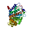


 PDBj
PDBj
 Assembly
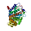


 Mass: 616.487 Da / Num. of mol.: 1 / Source method: obtained synthetically / Formula: C34H32FeN4O4
 Mass: 133.151 Da / Num. of mol.: 1 / Source method: obtained synthetically / Formula: C7H7N3
 Mass: 18.015 Da / Num. of mol.: 414 / Source method: isolated from a natural source / Formula: H2O
 Sample preparation
 / Beamline: 8.3.1 / Wavelength: 1.115869 Å
 Processing