 Movie
Movie Controller

[English] 日本語
 Yorodumi
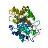
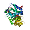
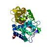
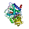
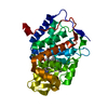
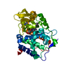
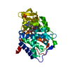
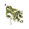
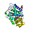
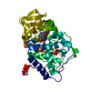
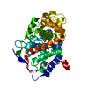
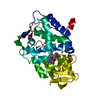
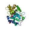
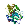
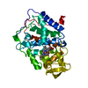
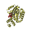
- PDB-1kxn: Crystal Structure of Cytochrome c Peroxidase with a Proposed Elec... -
+ Open data

- Basic information
| Entry | Database: PDB / ID: 1kxn | ||||||
|---|---|---|---|---|---|---|---|
| Title | Crystal Structure of Cytochrome c Peroxidase with a Proposed Electron Transfer Pathway Excised to Form a Ligand Binding Channel. | ||||||
 Components Components | cytochrome c peroxidase | ||||||
 Keywords Keywords | OXIDOREDUCTASE / engineered heme channel | ||||||
| Function / homology |  Function and homology information Function and homology informationcytochrome-c peroxidase / cytochrome-c peroxidase activity / hydrogen peroxide catabolic process / response to reactive oxygen species / peroxidase activity / mitochondrial intermembrane space / cellular response to oxidative stress / mitochondrial matrix / heme binding / mitochondrion / metal ion binding Similarity search - Function | ||||||
| Biological species |  | ||||||
| Method |  X-RAY DIFFRACTION / MOLECULAR REPLACEMENT / Resolution: 1.8 Å X-RAY DIFFRACTION / MOLECULAR REPLACEMENT / Resolution: 1.8 Å | ||||||
 Authors Authors | Rosenfeld, R.J. / Hayes, A.M.A. / Musah, R.A. / Goodin, D.B. | ||||||
 Citation Citation | Journal: Protein Sci. / Year: 2002 Title: Excision of a proposed electron transfer pathway in cytochrome c peroxidase and its replacement by a ligand-binding channel. Authors: Rosenfeld, R.J. / Hays, A.M. / Musah, R.A. / Goodin, D.B. | ||||||
| History |
|
- Structure visualization
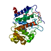
| Structure viewer | Molecule: MolmilJmol/JSmol |
|---|
- Downloads & links
-Download
| PDBx/mmCIF format | 1kxn.cif.gz | 82 KB | Display | PDBx/mmCIF format |
|---|---|---|---|---|
| PDB format | pdb1kxn.ent.gz | 59.2 KB | Display | PDB format |
| PDBx/mmJSON format | 1kxn.json.gz | Tree view | PDBx/mmJSON format | |
| Others |  Other downloads Other downloads |
-Validation report
| Arichive directory | https://data.pdbj.org/pub/pdb/validation_reports/kx/1kxnftp://data.pdbj.org/pub/pdb/validation_reports/kx/1kxn | HTTPS FTP |
|---|
-Related structure data
| Related structure data |  1kxmC 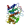 1cmqS S: Starting model for refinement C: citing same article ( |
|---|---|
| Similar structure data |


-Links
 PDBj
PDBj
- Assembly
| Deposited unit | 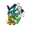
| ||||||||
|---|---|---|---|---|---|---|---|---|---|
| 1 |
| ||||||||
| Unit cell |
|
-Components
| #1: Protein | Mass: 32928.582 Da / Num. of mol.: 1 / Fragment: RESIDUES 72-362, NUMBERED 4-292 / Mutation: P190G Source method: isolated from a genetically manipulated source Source: (gene. exp.) Gene: CCP-MKT / Plasmid: PT7CCP / Species (production host): Escherichia coli / Production host:  |
|---|---|
| #2: Chemical | ChemComp-HEM /   Mass: 616.487 Da / Num. of mol.: 1 / Source method: obtained synthetically / Formula: C34H32FeN4O4 Mass: 616.487 Da / Num. of mol.: 1 / Source method: obtained synthetically / Formula: C34H32FeN4O4 |
| #3: Water | ChemComp-HOH /  Mass: 18.015 Da / Num. of mol.: 348 / Source method: isolated from a natural source / Formula: H2O Mass: 18.015 Da / Num. of mol.: 348 / Source method: isolated from a natural source / Formula: H2O |
-Experimental details
-Experiment
| Experiment | Method: X-RAY DIFFRACTION / Number of used crystals: 1 |
|---|
- Sample preparation
| Crystal | Density Matthews: 3.14 Å3/Da / Density % sol: 60.82 % | ||||||||||||||||||||||||||||||
|---|---|---|---|---|---|---|---|---|---|---|---|---|---|---|---|---|---|---|---|---|---|---|---|---|---|---|---|---|---|---|---|
| Crystal grow | Temperature: 298 K / Method: vapor diffusion, sitting drop / pH: 6 Details: MPD, pH 6.0, VAPOR DIFFUSION, SITTING DROP, temperature 298K | ||||||||||||||||||||||||||||||
| Crystal grow | *PLUS Temperature: 15 ℃ | ||||||||||||||||||||||||||||||
| Components of the solutions | *PLUS
|
-Data collection
| Diffraction | Mean temperature: 100 K |
|---|---|
| Diffraction source | Source: ROTATING ANODE / Type: RIGAKU / Wavelength: 1.54 Å |
| Detector | Type: SIEMENS / Detector: AREA DETECTOR / Date: Jan 15, 1996 |
| Radiation | Monochromator: graphite / Protocol: SINGLE WAVELENGTH / Monochromatic (M) / Laue (L): M / Scattering type: x-ray |
| Radiation wavelength | Wavelength: 1.54 Å / Relative weight: 1 |
| Reflection | Resolution: 1.8→10 Å / Num. obs: 35095 / % possible obs: 91.2 % / Observed criterion σ(F): 0 / Observed criterion σ(I): 0 / Redundancy: 4.6 % / Rsym value: 0.075 |
| Reflection shell | Resolution: 1.8→1.86 Å / Redundancy: 3.5 % / Num. unique all: 3208 / % possible all: 81.1 |
| Reflection | *PLUS Num. obs: 36741 / Num. measured all: 168657 / Rmerge(I) obs: 0.075 |
| Reflection shell | *PLUS Highest resolution: 1.8 Å / Lowest resolution: 1.85 Å / % possible obs: 81.1 % / Num. unique obs: 3208 / Num. measured obs: 11517 / Rmerge(I) obs: 0.55 / Mean I/σ(I) obs: 3.1 |
- Processing
| Software |
| |||||||||||||||||||||||||
|---|---|---|---|---|---|---|---|---|---|---|---|---|---|---|---|---|---|---|---|---|---|---|---|---|---|---|
| Refinement | Method to determine structure: MOLECULAR REPLACEMENT Starting model: PDB ENTRY 1CMQ Resolution: 1.8→10 Å / Num. parameters: 10827 / Num. restraintsaints: 9732 / σ(F): 0 / Stereochemistry target values: Engh & Huber
| |||||||||||||||||||||||||
| Solvent computation | Solvent model: MOEWS & KRETSINGER | |||||||||||||||||||||||||
| Refine analyze | Num. disordered residues: 0 / Occupancy sum hydrogen: 0 / Occupancy sum non hydrogen: 2706 | |||||||||||||||||||||||||
| Refinement step | Cycle: LAST / Resolution: 1.8→10 Å
| |||||||||||||||||||||||||
| Refine LS restraints |
| |||||||||||||||||||||||||
| Software | *PLUS Name: SHELXL-96 / Classification: refinement | |||||||||||||||||||||||||
| Refinement | *PLUS σ(F): 0 / Rfactor all: 0.1871 | |||||||||||||||||||||||||
| Solvent computation | *PLUS | |||||||||||||||||||||||||
| Displacement parameters | *PLUS Biso mean: 21.2 Å2 | |||||||||||||||||||||||||
| Refine LS restraints | *PLUS
|
