 Movie
Movie Controller

[English] 日本語
 Yorodumi
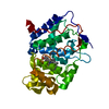
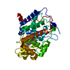
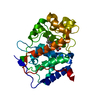
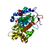
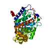
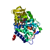
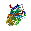
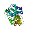
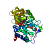
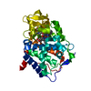
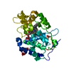
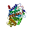
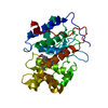
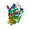
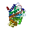
- PDB-4jm9: Crystal structure of Cytochrome C Peroxidase W191G-Gateless in co... -
+ Open data

- Basic information
| Entry | Database: PDB / ID: 4jm9 | ||||||
|---|---|---|---|---|---|---|---|
| Title | Crystal structure of Cytochrome C Peroxidase W191G-Gateless in complex with 3-amino-1-methylpyridinium | ||||||
 Components Components | Cytochrome c peroxidase | ||||||
 Keywords Keywords | OXIDOREDUCTASE / Model system / bulk solvent / ordered waters / docking / ligand binding / free energy calculation / molecular dynamics | ||||||
| Function / homology |  Function and homology information Function and homology informationcytochrome-c peroxidase activity / Oxidoreductases; Acting on a peroxide as acceptor; Peroxidases / hydrogen peroxide catabolic process / response to reactive oxygen species / mitochondrial intermembrane space / cellular response to oxidative stress / mitochondrial matrix / heme binding / metal ion binding Similarity search - Function | ||||||
| Biological species |  | ||||||
| Method |  X-RAY DIFFRACTION / SYNCHROTRON / MOLECULAR REPLACEMENT / Resolution: 1.41 Å X-RAY DIFFRACTION / SYNCHROTRON / MOLECULAR REPLACEMENT / Resolution: 1.41 Å | ||||||
 Authors Authors | Boyce, S.E. / Fischer, M. / Fish, I. | ||||||
 Citation Citation | Journal: J.Mol.Biol. / Year: 2013 Title: Blind prediction of charged ligand binding affinities in a model binding site. Authors: Rocklin, G.J. / Boyce, S.E. / Fischer, M. / Fish, I. / Mobley, D.L. / Shoichet, B.K. / Dill, K.A. #1: Journal: To be PublishedTitle: Docking to a water-filled model binding site in Cytochrome c Peroxidase Authors: Barelier, S. / Boyce, S.E. / Fischer, M. / Fish, I. / Goodin, D.B. / Shoichet, B.K. | ||||||
| History |
|
- Structure visualization
| Structure viewer | Molecule: MolmilJmol/JSmol |
|---|
- Downloads & links
-Download
| PDBx/mmCIF format | 4jm9.cif.gz | 173.8 KB | Display | PDBx/mmCIF format |
|---|---|---|---|---|
| PDB format | pdb4jm9.ent.gz | 138.5 KB | Display | PDB format |
| PDBx/mmJSON format | 4jm9.json.gz | Tree view | PDBx/mmJSON format | |
| Others |  Other downloads Other downloads |
-Validation report
| Arichive directory | https://data.pdbj.org/pub/pdb/validation_reports/jm/4jm9ftp://data.pdbj.org/pub/pdb/validation_reports/jm/4jm9 | HTTPS FTP |
|---|
-Related structure data
| Related structure data | 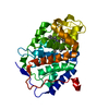 4jm5C 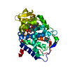 4jm6C 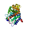 4jm8C 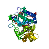 4jmaC 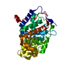 4jmwC  4jplC 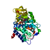 4jptC 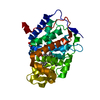 4jpuC 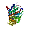 4jqjC 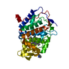 4jqkC  4jqmC 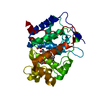 4jqnC C: citing same article ( |
|---|---|
| Similar structure data |


-Links
 PDBj
PDBj
- Assembly
| Deposited unit | 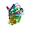
| ||||||||
|---|---|---|---|---|---|---|---|---|---|
| 1 |
| ||||||||
| Unit cell |
|
-Components
-Protein , 1 types, 1 molecules A
| #1: Protein | Mass: 32928.582 Da / Num. of mol.: 1 / Fragment: RESIDUES 72-362, deletions G192-A193 / Mutation: P190G, W191G Source method: isolated from a genetically manipulated source Source: (gene. exp.) Strain: RM11-1a / Gene: CCP1 CCP CPO YKR066C, SCRG_04081 / Production host:  |
|---|
-Non-polymers , 5 types, 439 molecules 


| #2: Chemical | ChemComp-HEM /  Mass: 616.487 Da / Num. of mol.: 1 / Source method: obtained synthetically / Formula: C34H32FeN4O4 Mass: 616.487 Da / Num. of mol.: 1 / Source method: obtained synthetically / Formula: C34H32FeN4O4 |
|---|---|
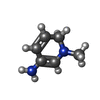
| #3: Chemical | ChemComp-LG5 /  Mass: 110.157 Da / Num. of mol.: 1 / Source method: obtained synthetically / Formula: C6H10N2 Mass: 110.157 Da / Num. of mol.: 1 / Source method: obtained synthetically / Formula: C6H10N2 |
| #4: Chemical | ChemComp-IOD /  Mass: 126.904 Da / Num. of mol.: 1 / Source method: obtained synthetically / Formula: I Mass: 126.904 Da / Num. of mol.: 1 / Source method: obtained synthetically / Formula: I |
| #5: Chemical | ChemComp-PO4 /  Mass: 94.971 Da / Num. of mol.: 1 / Source method: obtained synthetically / Formula: PO4 Mass: 94.971 Da / Num. of mol.: 1 / Source method: obtained synthetically / Formula: PO4 |
| #6: Water | ChemComp-HOH / Mass: 18.015 Da / Num. of mol.: 435 / Source method: isolated from a natural source / Formula: H2O |
-Experimental details
-Experiment
| Experiment | Method: X-RAY DIFFRACTION / Number of used crystals: 1 |
|---|
- Sample preparation
| Crystal | Density Matthews: 3.08 Å3/Da / Density % sol: 60.12 % |
|---|---|
| Crystal grow | Temperature: 283 K / Method: vapor diffusion, sitting drop / pH: 6 Details: Apo crystal grown in 500mM MES, pH 6.0. Soaked into 50mM 3-amino-1-methylpyridinium, 25% MPD pH 4.5 (30min-1h), vapor diffusion, sitting drop, temperature 283K |
-Data collection
| Diffraction | Mean temperature: 80 K |
|---|---|
| Diffraction source | Source: SYNCHROTRON / Site: ALS  / Beamline: 8.3.1 / Beamline: 8.3.1 |
| Detector | Type: ADSC QUANTUM 315r / Detector: CCD / Date: Jun 16, 2009 |
| Radiation | Monochromator: Double flat crystal, Si(111) / Protocol: SINGLE WAVELENGTH / Monochromatic (M) / Laue (L): M / Scattering type: x-ray |
| Radiation wavelength | Relative weight: 1 |
| Reflection | Resolution: 1.41→61.2 Å / Num. obs: 79143 |
- Processing
| Software |
| ||||||||||||||||||||||||||||||||||||||||||||||||||||||||||||||||||||||||||||||||||||||||||
|---|---|---|---|---|---|---|---|---|---|---|---|---|---|---|---|---|---|---|---|---|---|---|---|---|---|---|---|---|---|---|---|---|---|---|---|---|---|---|---|---|---|---|---|---|---|---|---|---|---|---|---|---|---|---|---|---|---|---|---|---|---|---|---|---|---|---|---|---|---|---|---|---|---|---|---|---|---|---|---|---|---|---|---|---|---|---|---|---|---|---|---|
| Refinement | Method to determine structure: MOLECULAR REPLACEMENT / Resolution: 1.41→29.16 Å / Cor.coef. Fo:Fc: 0.981 / Cor.coef. Fo:Fc free: 0.975 / Occupancy max: 1 / Occupancy min: 0.2 / SU B: 1.173 / SU ML: 0.022 / Cross valid method: THROUGHOUT / σ(F): 0 / ESU R: 0.046 / ESU R Free: 0.042 / Stereochemistry target values: MAXIMUM LIKELIHOOD Details: HYDROGENS HAVE BEEN ADDED IN THE RIDING POSITIONS U VALUES: REFINED INDIVIDUALLY
| ||||||||||||||||||||||||||||||||||||||||||||||||||||||||||||||||||||||||||||||||||||||||||
| Solvent computation | Ion probe radii: 0.8 Å / Shrinkage radii: 0.8 Å / VDW probe radii: 1.4 Å / Solvent model: BABINET MODEL WITH MASK | ||||||||||||||||||||||||||||||||||||||||||||||||||||||||||||||||||||||||||||||||||||||||||
| Displacement parameters | Biso max: 76.89 Å2 / Biso mean: 20.0436 Å2 / Biso min: 9.95 Å2
| ||||||||||||||||||||||||||||||||||||||||||||||||||||||||||||||||||||||||||||||||||||||||||
| Refinement step | Cycle: LAST / Resolution: 1.41→29.16 Å
| ||||||||||||||||||||||||||||||||||||||||||||||||||||||||||||||||||||||||||||||||||||||||||
| Refine LS restraints |
| ||||||||||||||||||||||||||||||||||||||||||||||||||||||||||||||||||||||||||||||||||||||||||
| LS refinement shell | Resolution: 1.41→1.444 Å / Total num. of bins used: 20
|
