 Movie
Movie Controller

[English] 日本語
 Yorodumi
- PDB-1aes: SPECIFICITY OF LIGAND BINDING TO A BURIED POLAR CAVITY AT THE ACT... -
+ Open data

- Basic information
| Entry | Database: PDB / ID: 1aes | ||||||
|---|---|---|---|---|---|---|---|
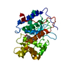
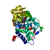
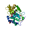
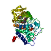
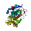
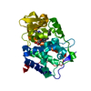
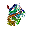
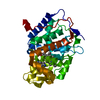
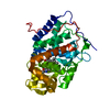
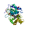
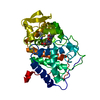
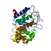
| Title | SPECIFICITY OF LIGAND BINDING TO A BURIED POLAR CAVITY AT THE ACTIVE SITE OF CYTOCHROME C PEROXIDASE (IMIDAZOLE) | ||||||
 Components Components | CYTOCHROME C PEROXIDASE | ||||||
 Keywords Keywords | OXIDOREDUCTASE / PEROXIDASE / TRANSIT PEPTIDE | ||||||
| Function / homology |  Function and homology information Function and homology informationcytochrome-c peroxidase / cytochrome-c peroxidase activity / hydrogen peroxide catabolic process / response to reactive oxygen species / peroxidase activity / mitochondrial intermembrane space / cellular response to oxidative stress / mitochondrial matrix / heme binding / mitochondrion / metal ion binding Similarity search - Function | ||||||
| Biological species |  | ||||||
| Method |  X-RAY DIFFRACTION / MOLECULAR REPLACEMENT / Resolution: 2.1 Å X-RAY DIFFRACTION / MOLECULAR REPLACEMENT / Resolution: 2.1 Å | ||||||
 Authors Authors | Musah, R.A. / Jensen, G.M. / Fitzgerald, M.M. / Mcree, D.E. / Goodin, D.B. | ||||||
 Citation Citation | Journal: Nat.Struct.Biol. / Year: 1996 Title: A ligand-gated, hinged loop rearrangement opens a channel to a buried artificial protein cavity. Authors: Fitzgerald, M.M. / Musah, R.A. / McRee, D.E. / Goodin, D.B. #1: Journal: Biochemistry / Year: 1994Title: Small Molecule Binding to an Artificially Created Cavity at the Active Site of Cytochrome C Peroxidase Authors: Fitzgerald, M.M. / Churchill, M.J. / Mcree, D.E. / Goodin, D.B. #2: Journal: Biochemistry / Year: 1993Title: The Asp-His-Fe Triad of Cytochrome C Peroxidase Controls the Reduction Potential, Electronic Structure, and Coupling of the Tryptophan Free Radical to the Heme Authors: Goodin, D.B. / Mcree, D.E. | ||||||
| History |
|
- Structure visualization
| Structure viewer | Molecule: MolmilJmol/JSmol |
|---|
- Downloads & links
-Download
| PDBx/mmCIF format | 1aes.cif.gz | 84.2 KB | Display | PDBx/mmCIF format |
|---|---|---|---|---|
| PDB format | pdb1aes.ent.gz | 63.6 KB | Display | PDB format |
| PDBx/mmJSON format | 1aes.json.gz | Tree view | PDBx/mmJSON format | |
| Others |  Other downloads Other downloads |
-Validation report
| Arichive directory | https://data.pdbj.org/pub/pdb/validation_reports/ae/1aesftp://data.pdbj.org/pub/pdb/validation_reports/ae/1aes | HTTPS FTP |
|---|
-Related structure data
| Related structure data | 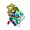 1aa4SC 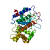 1aetC  1aeuC 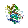 1cciC 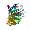 1rycC S: Starting model for refinement C: citing same article ( |
|---|---|
| Similar structure data |


-Links
 PDBj
PDBj


- Assembly
| Deposited unit | 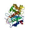
| ||||||||
|---|---|---|---|---|---|---|---|---|---|
| 1 |
| ||||||||
| Unit cell |
|
-Components
| #1: Protein | Mass: 33459.242 Da / Num. of mol.: 1 / Mutation: W191G Source method: isolated from a genetically manipulated source Details: CRYSTAL FORM BY Source: (gene. exp.) Cell line: BL21 / Cellular location: MITOCHONDRIA / Gene: CCP / Organelle: MITOCHONDRIA / Plasmid: PT7CCP / Species (production host): Escherichia coli / Cellular location (production host): CYTOPLASM / Gene (production host): CCP (MKT) / Production host:  |
|---|---|
| #2: Chemical | ChemComp-HEM /   Mass: 616.487 Da / Num. of mol.: 1 / Source method: obtained synthetically / Formula: C34H32FeN4O4 Mass: 616.487 Da / Num. of mol.: 1 / Source method: obtained synthetically / Formula: C34H32FeN4O4 |
| #3: Chemical | ChemComp-IMD /   Mass: 69.085 Da / Num. of mol.: 1 / Source method: obtained synthetically / Formula: C3H5N2 Mass: 69.085 Da / Num. of mol.: 1 / Source method: obtained synthetically / Formula: C3H5N2 |
| #4: Water | ChemComp-HOH /  Mass: 18.015 Da / Num. of mol.: 50 / Source method: isolated from a natural source / Formula: H2O Mass: 18.015 Da / Num. of mol.: 50 / Source method: isolated from a natural source / Formula: H2O |
-Experimental details
-Experiment
| Experiment | Method: X-RAY DIFFRACTION / Number of used crystals: 1 |
|---|
- Sample preparation
| Crystal | Density Matthews: 3.2 Å3/Da / Density % sol: 61 % | |||||||||||||||||||||||||
|---|---|---|---|---|---|---|---|---|---|---|---|---|---|---|---|---|---|---|---|---|---|---|---|---|---|---|
| Crystal grow | pH: 6 Details: 20% MPD, 40 MM PHOSPHATE PH 6.0. A SINGLE CRYSTAL OF W191G WAS SOAKED IN 50MM IMIDAZOLE IN 40% MPD AND 60 MM PHOSPHATE AT PH 4.5. | |||||||||||||||||||||||||
| Crystal grow | *PLUS Temperature: 4 ℃ / Method: vapor diffusion, sitting drop | |||||||||||||||||||||||||
| Components of the solutions | *PLUS
|
-Data collection
| Diffraction | Mean temperature: 290 K |
|---|---|
| Diffraction source | Source: ROTATING ANODE / Type: RIGAKU RUH2R / Wavelength: 1.5418 |
| Detector | Type: SIEMENS / Detector: AREA DETECTOR / Date: Jan 1, 1993 |
| Radiation | Monochromatic (M) / Laue (L): M / Scattering type: x-ray |
| Radiation wavelength | Wavelength: 1.5418 Å / Relative weight: 1 |
| Reflection | Resolution: 2.1→15 Å / Num. obs: 16612 / % possible obs: 64 % / Observed criterion σ(I): 0 / Redundancy: 3.73 % / Rmerge(I) obs: 0.087 / Rsym value: 0.077 / Net I/σ(I): 16.3 |
| Reflection shell | Resolution: 2.1→2.23 Å / Redundancy: 2.7 % / Rmerge(I) obs: 0.33 / Mean I/σ(I) obs: 0.702 / Rsym value: 0.62 / % possible all: 30 |
| Reflection | *PLUS Rmerge(I) obs: 0.087 |
- Processing
| Software |
| ||||||||||||
|---|---|---|---|---|---|---|---|---|---|---|---|---|---|
| Refinement | Method to determine structure: MOLECULAR REPLACEMENT Starting model: PDB ENTRY 1AA4 Resolution: 2.1→7 Å / Data cutoff high absF: 10000000 / Data cutoff low absF: 0.5 / σ(F): 0 /
| ||||||||||||
| Refinement step | Cycle: LAST / Resolution: 2.1→7 Å
|
