 Movie
Movie Controller

[English] 日本語
 Yorodumi
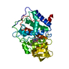
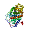
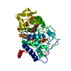
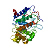
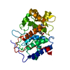
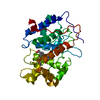
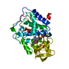
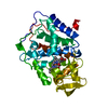
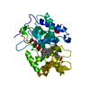
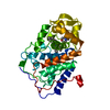
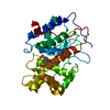
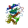
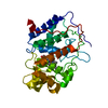
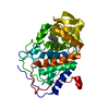
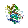
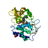
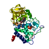
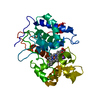
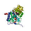
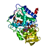
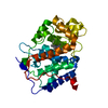
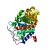
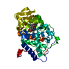
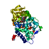
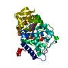
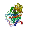
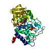
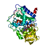
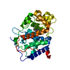
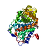
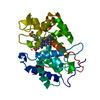
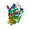
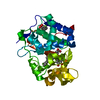
- PDB-2v2e: Structure of isoniazid (INH) bound to cytochrome c peroxidase mut... -
+ Open data

- Basic information
| Entry | Database: PDB / ID: 2v2e | ||||||
|---|---|---|---|---|---|---|---|
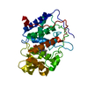
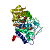
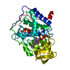
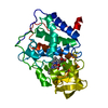
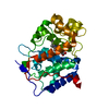
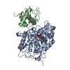
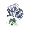
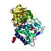
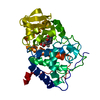
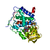
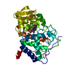
| Title | Structure of isoniazid (INH) bound to cytochrome c peroxidase mutant N184R Y36A | ||||||
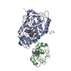
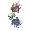
 Components Components | CYTOCHROME C PEROXIDASE | ||||||
 Keywords Keywords | OXIDOREDUCTASE / CYTOCHROME C PEROXIDASE / TRANSIT PEPTIDE / ORGANIC RADICAL / HYDROGEN PEROXIDE / INH / CCP / IRON / HEME / ISONIAZID / PEROXIDASE / MITOCHONDRION / METAL-BINDING | ||||||
| Function / homology |  Function and homology information Function and homology informationcytochrome-c peroxidase / cytochrome-c peroxidase activity / hydrogen peroxide catabolic process / response to reactive oxygen species / peroxidase activity / mitochondrial intermembrane space / cellular response to oxidative stress / mitochondrial matrix / heme binding / mitochondrion / metal ion binding Similarity search - Function | ||||||
| Biological species |  | ||||||
| Method |  X-RAY DIFFRACTION / MOLECULAR REPLACEMENT / Resolution: 1.68 Å X-RAY DIFFRACTION / MOLECULAR REPLACEMENT / Resolution: 1.68 Å | ||||||
 Authors Authors | Metcalfe, C.L. / Macdonald, I.K. / Brown, K.A. / Raven, E.L. / Moody, P.C.E. | ||||||
 Citation Citation | Journal: J.Biol.Chem. / Year: 2008 Title: The Tuberculosis Prodrug Isoniazid Bound to Activating Peroxidases. Authors: Metcalfe, C.L. / Macdonald, I.K. / Murphy, E.J. / Brown, K.A. / Raven, E.L. / Moody, P.C.E. | ||||||
| History |
|
- Structure visualization
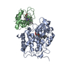
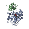
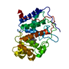
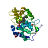
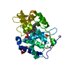
| Structure viewer | Molecule: MolmilJmol/JSmol |
|---|
- Downloads & links
-Download
| PDBx/mmCIF format | 2v2e.cif.gz | 143.2 KB | Display | PDBx/mmCIF format |
|---|---|---|---|---|
| PDB format | pdb2v2e.ent.gz | 110.7 KB | Display | PDB format |
| PDBx/mmJSON format | 2v2e.json.gz | Tree view | PDBx/mmJSON format | |
| Others |  Other downloads Other downloads |
-Validation report
| Arichive directory | https://data.pdbj.org/pub/pdb/validation_reports/v2/2v2eftp://data.pdbj.org/pub/pdb/validation_reports/v2/2v2e | HTTPS FTP |
|---|
-Related structure data
| Related structure data | 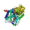 2v23SC  2vcfC 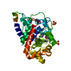 2vcnC 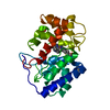 2vcsC S: Starting model for refinement C: citing same article ( |
|---|---|
| Similar structure data |


-Links
 PDBj
PDBj
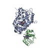
- Assembly
| Deposited unit | 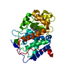
| ||||||||
|---|---|---|---|---|---|---|---|---|---|
| 1 |
| ||||||||
| Unit cell |
|
-Components
| #1: Protein | Mass: 33583.406 Da / Num. of mol.: 1 / Fragment: RESIDUES 71-361 / Mutation: YES Source method: isolated from a genetically manipulated source Source: (gene. exp.) Plasmid: PT7CCP/Y39A/N184R / Production host:  |
|---|---|
| #2: Chemical | ChemComp-HEM /   Mass: 616.487 Da / Num. of mol.: 1 / Source method: obtained synthetically / Formula: C34H32FeN4O4 Mass: 616.487 Da / Num. of mol.: 1 / Source method: obtained synthetically / Formula: C34H32FeN4O4 |
| #3: Chemical | ChemComp-ISZ / 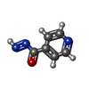  Mass: 135.123 Da / Num. of mol.: 1 / Source method: obtained synthetically / Formula: C6H5N3O Mass: 135.123 Da / Num. of mol.: 1 / Source method: obtained synthetically / Formula: C6H5N3O |
| #4: Water | ChemComp-HOH /  Mass: 18.015 Da / Num. of mol.: 315 / Source method: isolated from a natural source / Formula: H2O Mass: 18.015 Da / Num. of mol.: 315 / Source method: isolated from a natural source / Formula: H2O |
| Compound details | ENGINEERED |
-Experimental details
-Experiment
| Experiment | Method: X-RAY DIFFRACTION / Number of used crystals: 1 |
|---|
- Sample preparation
| Crystal | Density Matthews: 3.06 Å3/Da / Density % sol: 59.84 % / Description: NONE |
|---|---|
| Crystal grow | Method: microdialysis / pH: 6 Details: MICRODIALYSIS INTO 50MM POTASSIUM PHOSPHATE, PH 6.0 CONTAINING 30% V/V MPD. ISONIAZID WAS DISSOLVED INTO THE MOTHER LIQUOR (300MM) AND SOAKED INTO THE CRYSTAL. |
-Data collection
| Diffraction | Mean temperature: 100 K |
|---|---|
| Diffraction source | Source: ROTATING ANODE / Type: RIGAKU RUH2R / Wavelength: 1.5418 |
| Detector | Type: RIGAKU IMAGE PLATE / Detector: IMAGE PLATE / Details: XENOCS MULTI-LAYER |
| Radiation | Protocol: SINGLE WAVELENGTH / Monochromatic (M) / Laue (L): M / Scattering type: x-ray |
| Radiation wavelength | Wavelength: 1.5418 Å / Relative weight: 1 |
| Reflection | Resolution: 1.68→30.28 Å / Num. obs: 38990 / % possible obs: 81.9 % / Redundancy: 3.3 % / Rmerge(I) obs: 0.03 / Net I/σ(I): 27.4 |
| Reflection shell | Resolution: 1.68→1.77 Å / Redundancy: 1.2 % / Rmerge(I) obs: 0.13 / Mean I/σ(I) obs: 5.8 / % possible all: 20.3 |
- Processing
| Software |
| ||||||||||||||||||||||||||||||||||||||||||||||||||||||||||||||||||||||||||||||||||||||||||||||||||||||||||||||||||||||||||||||||||||||||||||||||||||||||||||||||||||||||||||||||||||||
|---|---|---|---|---|---|---|---|---|---|---|---|---|---|---|---|---|---|---|---|---|---|---|---|---|---|---|---|---|---|---|---|---|---|---|---|---|---|---|---|---|---|---|---|---|---|---|---|---|---|---|---|---|---|---|---|---|---|---|---|---|---|---|---|---|---|---|---|---|---|---|---|---|---|---|---|---|---|---|---|---|---|---|---|---|---|---|---|---|---|---|---|---|---|---|---|---|---|---|---|---|---|---|---|---|---|---|---|---|---|---|---|---|---|---|---|---|---|---|---|---|---|---|---|---|---|---|---|---|---|---|---|---|---|---|---|---|---|---|---|---|---|---|---|---|---|---|---|---|---|---|---|---|---|---|---|---|---|---|---|---|---|---|---|---|---|---|---|---|---|---|---|---|---|---|---|---|---|---|---|---|---|---|---|
| Refinement | Method to determine structure: MOLECULAR REPLACEMENT Starting model: PDB ENTRY 2V23 Resolution: 1.68→30.28 Å / Cor.coef. Fo:Fc: 0.955 / Cor.coef. Fo:Fc free: 0.944 / SU B: 3.047 / SU ML: 0.047 / Cross valid method: THROUGHOUT / ESU R: 0.14 / ESU R Free: 0.093 / Stereochemistry target values: MAXIMUM LIKELIHOOD / Details: HYDROGENS HAVE BEEN ADDED IN THE RIDING POSITIONS.
| ||||||||||||||||||||||||||||||||||||||||||||||||||||||||||||||||||||||||||||||||||||||||||||||||||||||||||||||||||||||||||||||||||||||||||||||||||||||||||||||||||||||||||||||||||||||
| Solvent computation | Ion probe radii: 0.8 Å / Shrinkage radii: 0.8 Å / VDW probe radii: 1.4 Å / Solvent model: MASK | ||||||||||||||||||||||||||||||||||||||||||||||||||||||||||||||||||||||||||||||||||||||||||||||||||||||||||||||||||||||||||||||||||||||||||||||||||||||||||||||||||||||||||||||||||||||
| Displacement parameters | Biso mean: 13.15 Å2
| ||||||||||||||||||||||||||||||||||||||||||||||||||||||||||||||||||||||||||||||||||||||||||||||||||||||||||||||||||||||||||||||||||||||||||||||||||||||||||||||||||||||||||||||||||||||
| Refinement step | Cycle: LAST / Resolution: 1.68→30.28 Å
| ||||||||||||||||||||||||||||||||||||||||||||||||||||||||||||||||||||||||||||||||||||||||||||||||||||||||||||||||||||||||||||||||||||||||||||||||||||||||||||||||||||||||||||||||||||||
| Refine LS restraints |
|