 Movie
Movie Controller

[English] 日本語
 Yorodumi
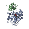
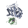
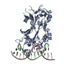
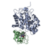
- PDB-2pcb: CRYSTAL STRUCTURE OF A COMPLEX BETWEEN ELECTRON TRANSFER PARTNERS... -
+ Open data

- Basic information
| Entry | Database: PDB / ID: 2pcb | ||||||
|---|---|---|---|---|---|---|---|
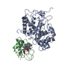
| Title | CRYSTAL STRUCTURE OF A COMPLEX BETWEEN ELECTRON TRANSFER PARTNERS, CYTOCHROME C PEROXIDASE AND CYTOCHROME C | ||||||
 Components Components |
| ||||||
 Keywords Keywords | OXIDOREDUCTASE/ELECTRON TRANSPORT / OXIDOREDUCTASE-ELECTRON TRANSPORT complex | ||||||
| Function / homology |  Function and homology information Function and homology informationcytochrome complex / cytochrome-c peroxidase / cytochrome-c peroxidase activity / mitochondrial electron transport, cytochrome c to oxygen / mitochondrial electron transport, ubiquinol to cytochrome c / hydrogen peroxide catabolic process / response to reactive oxygen species / peroxidase activity / apoptotic signaling pathway / mitochondrial intermembrane space ...cytochrome complex / cytochrome-c peroxidase / cytochrome-c peroxidase activity / mitochondrial electron transport, cytochrome c to oxygen / mitochondrial electron transport, ubiquinol to cytochrome c / hydrogen peroxide catabolic process / response to reactive oxygen species / peroxidase activity / apoptotic signaling pathway / mitochondrial intermembrane space / cellular response to oxidative stress / electron transfer activity / mitochondrial matrix / heme binding / lipid binding / mitochondrion / metal ion binding / identical protein binding / cytosol Similarity search - Function | ||||||
| Biological species |   | ||||||
| Method |  X-RAY DIFFRACTION / Resolution: 2.8 Å X-RAY DIFFRACTION / Resolution: 2.8 Å | ||||||
 Authors Authors | Pelletier, H. / Kraut, J. | ||||||
 Citation Citation | Journal: Science / Year: 1992 Title: Crystal structure of a complex between electron transfer partners, cytochrome c peroxidase and cytochrome c. Authors: Pelletier, H. / Kraut, J. #1: Journal: Biochemistry / Year: 1990Title: X-Ray Structures of Recombinant Yeast Cytochrome C Peroxidase and Three Heme-Cleft Mutants Prepared by Site-Directed Mutagenesis Authors: Wang, J. / Mauro, J.M. / Edwards, S.L. / Oatley, S.J. / Fishel, L.A. / Ashford, V.A. / Xuong, N.-H. / Kraut, J. #2: Journal: J.Mol.Biol. / Year: 1990Title: High-Resolution Three Dimensional Structure of Horse Heart Cytochrome C Authors: Bushnell, G.W. / Louie, G.V. / Brayer, G.D. #3: Journal: J.Biol.Chem. / Year: 1987Title: Co-Crystals of Yeast Cytochrome C Peroxidase and Horse Heart Cytochrome C Authors: Poulos, T.L. / Sheriff, S. / Howard, A.J. | ||||||
| History |
|
- Structure visualization
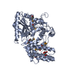
| Structure viewer | Molecule: MolmilJmol/JSmol |
|---|
- Downloads & links
-Download
| PDBx/mmCIF format | 2pcb.cif.gz | 162 KB | Display | PDBx/mmCIF format |
|---|---|---|---|---|
| PDB format | pdb2pcb.ent.gz | 127.5 KB | Display | PDB format |
| PDBx/mmJSON format | 2pcb.json.gz | Tree view | PDBx/mmJSON format | |
| Others |  Other downloads Other downloads |
-Validation report
| Arichive directory | https://data.pdbj.org/pub/pdb/validation_reports/pc/2pcbftp://data.pdbj.org/pub/pdb/validation_reports/pc/2pcb | HTTPS FTP |
|---|
-Related structure data


-Links
 PDBj
PDBj


- Assembly
| Deposited unit | 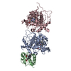
| ||||||||
|---|---|---|---|---|---|---|---|---|---|
| 1 | 
| ||||||||
| 2 | 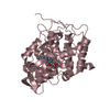
| ||||||||
| Unit cell |
|
-Components
| #1: Protein | Mass: 33769.605 Da / Num. of mol.: 2 Source method: isolated from a genetically manipulated source Source: (gene. exp.) Organ: HEART / References: UniProt: P00431 #2: Protein | | Mass: 11725.598 Da / Num. of mol.: 1 Source method: isolated from a genetically manipulated source Source: (gene. exp.) #3: Chemical |   Mass: 616.487 Da / Num. of mol.: 3 / Source method: obtained synthetically / Formula: C34H32FeN4O4 Mass: 616.487 Da / Num. of mol.: 3 / Source method: obtained synthetically / Formula: C34H32FeN4O4#4: Water | ChemComp-HOH / |  Mass: 18.015 Da / Num. of mol.: 337 / Source method: isolated from a natural source / Formula: H2O Mass: 18.015 Da / Num. of mol.: 337 / Source method: isolated from a natural source / Formula: H2O |
|---|
-Experimental details
-Experiment
| Experiment | Method: X-RAY DIFFRACTION |
|---|
- Sample preparation
| Crystal | Density Matthews: 3.25 Å3/Da / Density % sol: 62.2 % | ||||||||||||||||||||||||
|---|---|---|---|---|---|---|---|---|---|---|---|---|---|---|---|---|---|---|---|---|---|---|---|---|---|
| Crystal grow | *PLUS pH: 7 / Method: vapor diffusion, sitting drop | ||||||||||||||||||||||||
| Components of the solutions | *PLUS
|
-Data collection
| Reflection | *PLUS Highest resolution: 2.8 Å / Num. obs: 24671 / % possible obs: 93 % / Rmerge(I) obs: 0.055 |
|---|
- Processing
| Software | Name: X-PLOR / Classification: refinement | ||||||||||||
|---|---|---|---|---|---|---|---|---|---|---|---|---|---|
| Refinement | Resolution: 2.8→6 Å / Rfactor Rwork: 0.178 Details: THE YEAST CCP USED HERE IS A RECOMBINANT (CALLED CCP(MI) AND EXPRESSED IN E. COLI) WHICH HAS A MET-ILE DIPEPTIDE FUSED TO THE N-TERMINUS. THIS MET-ILE DIPEPTIDE WAS NOT INCLUDED IN THIS ...Details: THE YEAST CCP USED HERE IS A RECOMBINANT (CALLED CCP(MI) AND EXPRESSED IN E. COLI) WHICH HAS A MET-ILE DIPEPTIDE FUSED TO THE N-TERMINUS. THIS MET-ILE DIPEPTIDE WAS NOT INCLUDED IN THIS STRUCTURE DUE TO DISORDER. AFTER FINAL REFINEMENT THERE WAS ADDITIONAL ELECTRON DENSITY FOUND IN THE ASYMMETRIC UNIT WHICH HAS BEEN ATTRIBUTED TO A PARTIALLY OCCUPIED SECOND CYTOCHROME C MOLECULE. THE COORDINATES FOR A POSSIBLE SECOND CYTOCHROME C SITE HAVE NOT BEEN INCLUDED IN THIS ENTRY. SEE REFERENCE 1 FOR DETAILS. | ||||||||||||
| Refinement step | Cycle: LAST / Resolution: 2.8→6 Å
| ||||||||||||
| Refinement | *PLUS Highest resolution: 2.8 Å / Rfactor obs: 0.178 | ||||||||||||
| Solvent computation | *PLUS | ||||||||||||
| Displacement parameters | *PLUS | ||||||||||||
| Refine LS restraints | *PLUS
|
