 Movie
Movie Controller

[English] 日本語
 Yorodumi
- PDB-2pcc: CRYSTAL STRUCTURE OF A COMPLEX BETWEEN ELECTRON TRANSFER PARTNERS... -
+ Open data

- Basic information
| Entry | Database: PDB / ID: 2pcc | ||||||
|---|---|---|---|---|---|---|---|
| Title | CRYSTAL STRUCTURE OF A COMPLEX BETWEEN ELECTRON TRANSFER PARTNERS, CYTOCHROME C PEROXIDASE AND CYTOCHROME C | ||||||
 Components Components |
| ||||||
 Keywords Keywords | OXIDOREDUCTASE/ELECTRON TRANSPORT / OXIDOREDUCTASE-ELECTRON TRANSPORT complex | ||||||
| Function / homology |  Function and homology information Function and homology informationRelease of apoptotic factors from the mitochondria / Pyroptosis / Detoxification of Reactive Oxygen Species / Respiratory electron transport / cytochrome-c peroxidase / cytochrome-c peroxidase activity / cardiolipin binding / mitochondrial electron transport, cytochrome c to oxygen / mitochondrial electron transport, ubiquinol to cytochrome c / hydrogen peroxide catabolic process ...Release of apoptotic factors from the mitochondria / Pyroptosis / Detoxification of Reactive Oxygen Species / Respiratory electron transport / cytochrome-c peroxidase / cytochrome-c peroxidase activity / cardiolipin binding / mitochondrial electron transport, cytochrome c to oxygen / mitochondrial electron transport, ubiquinol to cytochrome c / hydrogen peroxide catabolic process / response to reactive oxygen species / peroxidase activity / mitochondrial intermembrane space / cellular response to oxidative stress / electron transfer activity / mitochondrial matrix / heme binding / mitochondrion / metal ion binding Similarity search - Function | ||||||
| Biological species |  | ||||||
| Method |  X-RAY DIFFRACTION / Resolution: 2.3 Å X-RAY DIFFRACTION / Resolution: 2.3 Å | ||||||
 Authors Authors | Pelletier, H. / Kraut, J. | ||||||
 Citation Citation | Journal: Science / Year: 1992 Title: Crystal structure of a complex between electron transfer partners, cytochrome c peroxidase and cytochrome c. Authors: Pelletier, H. / Kraut, J. #1: Journal: Biochemistry / Year: 1990Title: X-Ray Structures of Recombinant Yeast Cytochrome C Peroxidase and Three Heme-Cleft Mutants Prepared by Site-Directed Mutagenesis Authors: Wang, J. / Mauro, J.M. / Edwards, S.L. / Oatley, S.J. / Fishel, L.A. / Ashford, V.A. / Xuong, N.-H. / Kraut, J. #2: Journal: J.Mol.Biol. / Year: 1990Title: High-Resolution Refinement of Yeast Iso-1-Cytochrome C and Comparisons with Other Eukaryotic Cytochromes C Authors: Louie, G.V. / Brayer, G.D. | ||||||
| History |
|
- Structure visualization
| Structure viewer | Molecule: MolmilJmol/JSmol |
|---|
- Downloads & links
-Download
| PDBx/mmCIF format | 2pcc.cif.gz | 188.4 KB | Display | PDBx/mmCIF format |
|---|---|---|---|---|
| PDB format | pdb2pcc.ent.gz | 148.8 KB | Display | PDB format |
| PDBx/mmJSON format | 2pcc.json.gz | Tree view | PDBx/mmJSON format | |
| Others |  Other downloads Other downloads |
-Validation report
| Arichive directory | https://data.pdbj.org/pub/pdb/validation_reports/pc/2pccftp://data.pdbj.org/pub/pdb/validation_reports/pc/2pcc | HTTPS FTP |
|---|
-Related structure data


-Links
 PDBj
PDBj


- Assembly
| Deposited unit | 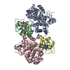
| ||||||||
|---|---|---|---|---|---|---|---|---|---|
| 1 | 
| ||||||||
| 2 | 
| ||||||||
| Unit cell |
| ||||||||
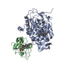
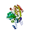
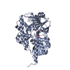
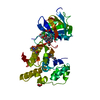
| Details | CHAINS LABELED A (CCP NUMBER 1) AND B (CYTOCHROME C NUMBER 1) REPRESENT ONE COMPLEX MOLECULE IN THE ASYMMETRIC UNIT, AND, LIKEWISE, CHAINS LABELED C (CCP NUMBER 2) AND D (CYTOCHROME C NUMBER 2) REPRESENT THE SECOND COMPLEX MOLECULE IN THE ASYMMETRIC UNIT. WATER MOLECULES NUMBERED 301 TO 554 ARE ASSOCIATED WITH THE FIRST COMPLEX, AND WATER MOLECULES NUMBERED 600 TO 848 ARE ASSOCIATED WITH THE SECOND COMPLEX. |
-Components
| #1: Protein | Mass: 33769.605 Da / Num. of mol.: 2 Source method: isolated from a genetically manipulated source Source: (gene. exp.) References: UniProt: P00431 #2: Protein | Mass: 12073.835 Da / Num. of mol.: 2 Source method: isolated from a genetically manipulated source Source: (gene. exp.) References: UniProt: P00044 #3: Chemical |   Mass: 96.063 Da / Num. of mol.: 2 / Source method: obtained synthetically / Formula: SO4 Mass: 96.063 Da / Num. of mol.: 2 / Source method: obtained synthetically / Formula: SO4#4: Chemical | ChemComp-HEM /   Mass: 616.487 Da / Num. of mol.: 4 / Source method: obtained synthetically / Formula: C34H32FeN4O4 Mass: 616.487 Da / Num. of mol.: 4 / Source method: obtained synthetically / Formula: C34H32FeN4O4#5: Water | ChemComp-HOH / |  Mass: 18.015 Da / Num. of mol.: 503 / Source method: isolated from a natural source / Formula: H2O Mass: 18.015 Da / Num. of mol.: 503 / Source method: isolated from a natural source / Formula: H2OSequence details | IN ACCORDANCE WITH THE SEQUENCE NUMBERING FOR EUKARYOTIC CYTOCHROMES C, THE SEQUENCE NUMBERING FOR ...IN ACCORDANCE | |
|---|
-Experimental details
-Experiment
| Experiment | Method: X-RAY DIFFRACTION |
|---|
- Sample preparation
| Crystal | Density Matthews: 2.49 Å3/Da / Density % sol: 50.69 % | ||||||||||||||||||||||||||||||||||||
|---|---|---|---|---|---|---|---|---|---|---|---|---|---|---|---|---|---|---|---|---|---|---|---|---|---|---|---|---|---|---|---|---|---|---|---|---|---|
| Crystal grow | *PLUS pH: 7 / Method: vapor diffusion, sitting drop | ||||||||||||||||||||||||||||||||||||
| Components of the solutions | *PLUS
|
-Data collection
| Reflection | *PLUS Highest resolution: 2.3 Å / Num. obs: 37845 / % possible obs: 93 % / Rmerge(I) obs: 0.044 |
|---|
- Processing
| Software |
| ||||||||||||
|---|---|---|---|---|---|---|---|---|---|---|---|---|---|
| Refinement | Resolution: 2.3→6 Å / Rfactor Rwork: 0.167 Details: THE YEAST CCP USED HERE IS A RECOMBINANT [CALLED CCP(MI) AND EXPRESSED IN E. COLI] WHICH HAS A MET-ILE DIPEPTIDE FUSED TO THE N-TERMINUS. THIS MET-ILE DIPEPTIDE WAS NOT INCLUDED IN THIS ...Details: THE YEAST CCP USED HERE IS A RECOMBINANT [CALLED CCP(MI) AND EXPRESSED IN E. COLI] WHICH HAS A MET-ILE DIPEPTIDE FUSED TO THE N-TERMINUS. THIS MET-ILE DIPEPTIDE WAS NOT INCLUDED IN THIS STRUCTURE DUE TO DISORDER. THERE WAS UNEXPLAINED ELECTRON DENSITY ABOVE THE HEME IRON INTO WHICH WATER MOLECULES ALONE COULD NOT BE MODELED. AN SO- GROUP, A POSSIBLE DEGRADATION PRODUCT OF DTT, WAS PLACED HERE AND WAS TREATED AS AN INCOMPLETE SULFATE ION DURING REFINEMENT. THIS IS NOT A SULFATE ION. IT IS ONLY CALLED A SULFATE ION FOR SIMPLICITY. | ||||||||||||
| Refinement step | Cycle: LAST / Resolution: 2.3→6 Å
| ||||||||||||
| Refinement | *PLUS Highest resolution: 2.3 Å / Rfactor obs: 0.172 | ||||||||||||
| Solvent computation | *PLUS | ||||||||||||
| Displacement parameters | *PLUS | ||||||||||||
| Refine LS restraints | *PLUS
|
