 Movie
Movie Controller

[English] 日本語
 Yorodumi
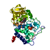
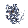
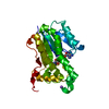
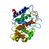
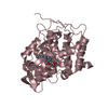
- PDB-1cyf: IDENTIFYING THE PHYSIOLOGICAL ELECTRON TRANSFER SITE OF CYTOCHROM... -
+ Open data

- Basic information
| Entry | Database: PDB / ID: 1cyf | ||||||
|---|---|---|---|---|---|---|---|
| Title | IDENTIFYING THE PHYSIOLOGICAL ELECTRON TRANSFER SITE OF CYTOCHROME C PEROXIDASE BY STRUCTURE-BASED ENGINEERING | ||||||
 Components Components | CYTOCHROME C PEROXIDASE | ||||||
 Keywords Keywords | OXIDOREDUCTASE (H2O2(A)) | ||||||
| Function / homology |  Function and homology information Function and homology informationcytochrome-c peroxidase / cytochrome-c peroxidase activity / hydrogen peroxide catabolic process / response to reactive oxygen species / peroxidase activity / mitochondrial intermembrane space / cellular response to oxidative stress / mitochondrial matrix / heme binding / mitochondrion / metal ion binding Similarity search - Function | ||||||
| Biological species |  | ||||||
| Method |  X-RAY DIFFRACTION / Resolution: 2.35 Å X-RAY DIFFRACTION / Resolution: 2.35 Å | ||||||
 Authors Authors | Miller, M.A. / Han, G.W. / Kraut, J. | ||||||
 Citation Citation | Journal: Biochemistry / Year: 1996 Title: Identifying the physiological electron transfer site of cytochrome c peroxidase by structure-based engineering. Authors: Miller, M.A. / Geren, L. / Han, G.W. / Saunders, A. / Beasley, J. / Pielak, G.J. / Durham, B. / Millett, F. / Kraut, J. #1: Journal: Biochemistry / Year: 1990Title: X-Ray Structures of Recombinant Yeast Cytochrome C Peroxidase and Three Heme-Cleft Mutants Prepared by Site-Directed Mutagenesis Authors: Wang, J. / Mauro, J.M. / Edwards, S.L. / Oatley, S.J. / Fishel, L.A. / Ashford, V.A. / Xuong, N.-H. / Kraut, J. #2: Journal: J.Biol.Chem. / Year: 1984Title: Crystal Structure of Yeast Cytochrome C Peroxidase Refined at 1.7 Angstroms Resolution Authors: Finzel, B.C. / Poulos, T.L. / Kraut, J. | ||||||
| History |
|
- Structure visualization
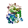
| Structure viewer | Molecule: MolmilJmol/JSmol |
|---|
- Downloads & links
-Download
| PDBx/mmCIF format | 1cyf.cif.gz | 77.9 KB | Display | PDBx/mmCIF format |
|---|---|---|---|---|
| PDB format | pdb1cyf.ent.gz | 57.9 KB | Display | PDB format |
| PDBx/mmJSON format | 1cyf.json.gz | Tree view | PDBx/mmJSON format | |
| Others |  Other downloads Other downloads |
-Validation report
| Arichive directory | https://data.pdbj.org/pub/pdb/validation_reports/cy/1cyfftp://data.pdbj.org/pub/pdb/validation_reports/cy/1cyf | HTTPS FTP |
|---|
-Related structure data
| Related structure data | |
|---|---|
| Similar structure data |


-Links
 PDBj
PDBj
- Assembly
| Deposited unit | 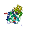
| ||||||||
|---|---|---|---|---|---|---|---|---|---|
| 1 |
| ||||||||
| Unit cell |
|
-Components
| #1: Protein | Mass: 33769.605 Da / Num. of mol.: 1 / Mutation: INS(MET ILE AT N-TERMINUS), C128A, A193C Source method: isolated from a genetically manipulated source Source: (gene. exp.) Production host:  |
|---|---|
| #2: Chemical | ChemComp-HEM /   Mass: 616.487 Da / Num. of mol.: 1 / Source method: obtained synthetically / Formula: C34H32FeN4O4 Mass: 616.487 Da / Num. of mol.: 1 / Source method: obtained synthetically / Formula: C34H32FeN4O4 |
| #3: Water | ChemComp-HOH /  Mass: 18.015 Da / Num. of mol.: 152 / Source method: isolated from a natural source / Formula: H2O Mass: 18.015 Da / Num. of mol.: 152 / Source method: isolated from a natural source / Formula: H2O |
| Compound details | THE SIDE CHAIN AT CYS 193 IS DISORDERED, AND NO INTERPRETABLE DENSITY WAS FOUND BEYOND SG OF CYS ...THE SIDE CHAIN AT CYS 193 IS DISORDERED |
| Sequence details | THIS CYTOCHROME C PEROXIDASE DIFFERS FROM A PREVIOUSLY DEPOSITED STRUCTURE (PROTEIN DATA BANK ENTRY ...THIS CYTOCHROME |
-Experimental details
-Experiment
| Experiment | Method: X-RAY DIFFRACTION |
|---|
- Sample preparation
| Crystal | Density Matthews: 2.49 Å3/Da / Density % sol: 50.66 % |
|---|---|
| Crystal grow | *PLUS Method: unknown |
-Data collection
| Radiation | Scattering type: x-ray |
|---|---|
| Radiation wavelength | Relative weight: 1 |
- Processing
| Software | Name: TNT / Classification: refinement | ||||||||||||||||||||||||||||||||||||||||||||||||||
|---|---|---|---|---|---|---|---|---|---|---|---|---|---|---|---|---|---|---|---|---|---|---|---|---|---|---|---|---|---|---|---|---|---|---|---|---|---|---|---|---|---|---|---|---|---|---|---|---|---|---|---|
| Refinement | Resolution: 2.35→20 Å / Isotropic thermal model: 0 / σ(F): 0 Stereochemistry target values: D.E. TRONRUD ET AL., ACTA CRYST. A43, 489 (1987) Details: THE ADDITION OF A BULKY GROUP AT POSITION 193 MAKES IT IMPOSSIBLE FOR THE ENZYME TO CRYSTALLIZE IN A FORM THAT IS ISOMORPHOUS WITH THE CCP(MI) PARENT. THIS IS BECAUSE CB OF ALA 193 IN THE ...Details: THE ADDITION OF A BULKY GROUP AT POSITION 193 MAKES IT IMPOSSIBLE FOR THE ENZYME TO CRYSTALLIZE IN A FORM THAT IS ISOMORPHOUS WITH THE CCP(MI) PARENT. THIS IS BECAUSE CB OF ALA 193 IN THE PARENT MAKES A CRYSTAL CONTACT WITH THE SIDE CHAIN OF THR 275. THE MUTANT ENZYME ADOPTS A NEW CRYSTAL PACKING THAT ALLOWS RESIDUE 193 TO PROJECT INTO AN OTHERWISE UNOCCUPIED AREA. THE MODEL WAS CREATED INITIALLY USING MOLECULAR REPLACEMENT WITH THE CCP(MI) PARENT AS THE SEARCH MODEL. TRANSLATION FUNCTIONS WERE CALCULATED BY SAMPLING A TRANSLATION VECTOR ON A 1 ANGSTROM GRID, UTILIZING DATA BETWEEN 8 AND 4 ANGSTROM RESOLUTION. THE TRANSLATION VECTOR WAS FURTHER REFINED BY AN R-FACTOR MINIMIZATION PROCEDURE WITH STEPS OF 0.1 ANGSTROM, RESULTING IN AN R-FACTOR OF 0.283 FOR DATA TO 4 ANGSTROMS RESOLUTION. UNLIKE THE CCP(MI) PARENT, COORDINATES FOR RESIDUES -1, 0, AND 1 - 3 ARE INCLUDED IN THIS ENTRY BECAUSE THESE RESIDUES COULD BE RESOLVED IN THE FINAL ELECTRON DENSITY MAPS. WATER MOLECULES WITH B-FACTORS GREATER THAN 80. WERE NOT INCLUDED.
| ||||||||||||||||||||||||||||||||||||||||||||||||||
| Solvent computation | Solvent model: MOEW & KRETSINGER, JMB 91, 211-228 (1975) / Bsol: 91.17 Å2 / ksol: 0.67 e/Å3 | ||||||||||||||||||||||||||||||||||||||||||||||||||
| Refinement step | Cycle: LAST / Resolution: 2.35→20 Å
| ||||||||||||||||||||||||||||||||||||||||||||||||||
| Refine LS restraints |
|
