 Movie
Movie Controller


+ Open data

- Basic information
| Entry | Database: PDB / ID: 1bva | ||||||
|---|---|---|---|---|---|---|---|
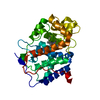
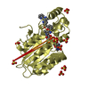
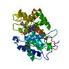
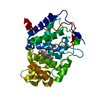
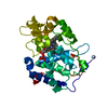
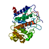
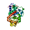
| Title | MANGANESE BINDING MUTANT IN CYTOCHROME C PEROXIDASE | ||||||
 Components Components | PROTEIN (CYTOCHROME C PEROXIDASE) | ||||||
 Keywords Keywords | OXIDOREDUCTASE / PEROXIDASE / METALLOENZYME / PROTEIN ENGINEERING | ||||||
| Function / homology |  Function and homology information Function and homology informationcytochrome-c peroxidase / cytochrome-c peroxidase activity / hydrogen peroxide catabolic process / response to reactive oxygen species / peroxidase activity / mitochondrial intermembrane space / cellular response to oxidative stress / mitochondrial matrix / heme binding / mitochondrion / metal ion binding Similarity search - Function | ||||||
| Biological species |  | ||||||
| Method |  X-RAY DIFFRACTION / MOLECULAR REPLACEMENT / Resolution: 1.89 Å X-RAY DIFFRACTION / MOLECULAR REPLACEMENT / Resolution: 1.89 Å | ||||||
 Authors Authors | Wilcox, S.K. / Mcree, D.E. / Goodin, D.B. | ||||||
 Citation Citation | Journal: Biochemistry / Year: 1998 Title: Rational design of a functional metalloenzyme: introduction of a site for manganese binding and oxidation into a heme peroxidase. Authors: Wilcox, S.K. / Putnam, C.D. / Sastry, M. / Blankenship, J. / Chazin, W.J. / McRee, D.E. / Goodin, D.B. | ||||||
| History |
|
- Structure visualization
| Structure viewer | Molecule: MolmilJmol/JSmol |
|---|
- Downloads & links
-Download
| PDBx/mmCIF format | 1bva.cif.gz | 83.8 KB | Display | PDBx/mmCIF format |
|---|---|---|---|---|
| PDB format | pdb1bva.ent.gz | 60.5 KB | Display | PDB format |
| PDBx/mmJSON format | 1bva.json.gz | Tree view | PDBx/mmJSON format | |
| Others |  Other downloads Other downloads |
-Validation report
| Arichive directory | https://data.pdbj.org/pub/pdb/validation_reports/bv/1bvaftp://data.pdbj.org/pub/pdb/validation_reports/bv/1bva | HTTPS FTP |
|---|
-Related structure data
| Related structure data | 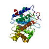 1ccgS S: Starting model for refinement |
|---|---|
| Similar structure data |


-Links
 PDBj
PDBj
- Assembly
| Deposited unit | 
| ||||||||
|---|---|---|---|---|---|---|---|---|---|
| 1 |
| ||||||||
| Unit cell |
|
-Components
| #1: Protein | Mass: 33635.371 Da / Num. of mol.: 1 / Mutation: D37E,P44D,V45D Source method: isolated from a genetically manipulated source Details: CRYSTAL FORM MKTBY Source: (gene. exp.) Strain: BL21 (DE3) / Description: MUTATION TO BIND MANGANESE / Cellular location: CYTOPLASM / Gene: CCP (MKT) / Organelle: MITOCHONDRIA / Plasmid: PT7CCP / Species (production host): Escherichia coli / Gene (production host): CCP (MKT) / Production host:  |
|---|---|
| #2: Chemical | ChemComp-MN /   Mass: 54.938 Da / Num. of mol.: 1 / Source method: obtained synthetically / Formula: Mn Mass: 54.938 Da / Num. of mol.: 1 / Source method: obtained synthetically / Formula: Mn |
| #3: Chemical | ChemComp-HEM /   Mass: 616.487 Da / Num. of mol.: 1 / Source method: obtained synthetically / Formula: C34H32FeN4O4 Mass: 616.487 Da / Num. of mol.: 1 / Source method: obtained synthetically / Formula: C34H32FeN4O4 |
| #4: Water | ChemComp-HOH /  Mass: 18.015 Da / Num. of mol.: 385 / Source method: isolated from a natural source / Formula: H2O Mass: 18.015 Da / Num. of mol.: 385 / Source method: isolated from a natural source / Formula: H2O |
| Sequence details | THIS CYTOCHROME C PEROXIDASE DIFFERS FROM 2CYP BY STRAIN RELATED SUBSTITUTIONS OF THR 53 WITH ILE, ...THIS CYTOCHROME |
-Experimental details
-Experiment
| Experiment | Method: X-RAY DIFFRACTION / Number of used crystals: 1 |
|---|
- Sample preparation
| Crystal | Density Matthews: 3.2 Å3/Da / Density % sol: 61 % Description: A 100K STRUCTURE OF 1CCG WAS USED INITIALLY BECAUSE IT HAD THE MOST SIMILAR UNIT CELL. THE APPROPRIATE AMINO ACID IDENTITIES WERE CHANGED. | ||||||||||||||||||||||||||||||||||||
|---|---|---|---|---|---|---|---|---|---|---|---|---|---|---|---|---|---|---|---|---|---|---|---|---|---|---|---|---|---|---|---|---|---|---|---|---|---|
| Crystal grow | pH: 6 / Details: 20% MPD, 35 MM PHOSPHATE, PH 6.0, 5 MM MNSO4. | ||||||||||||||||||||||||||||||||||||
| Crystal | *PLUS | ||||||||||||||||||||||||||||||||||||
| Crystal grow | *PLUS pH: 6 / Method: vapor diffusion, sitting drop | ||||||||||||||||||||||||||||||||||||
| Components of the solutions | *PLUS
|
-Data collection
| Diffraction | Mean temperature: 100 K |
|---|---|
| Diffraction source | Source: ROTATING ANODE / Type: SIEMENS / Wavelength: 1.5418 |
| Detector | Type: SIEMENS / Detector: AREA DETECTOR / Date: Jan 15, 1998 |
| Radiation | Protocol: SINGLE WAVELENGTH / Monochromatic (M) / Laue (L): M / Scattering type: x-ray |
| Radiation wavelength | Wavelength: 1.5418 Å / Relative weight: 1 |
| Reflection | Resolution: 1.88→15 Å / Num. obs: 26232 / % possible obs: 81 % / Observed criterion σ(I): 0 / Redundancy: 2.3 % / Rmerge(I) obs: 0.052 / Rsym value: 0.052 / Net I/σ(I): 18.8 |
| Reflection shell | Resolution: 1.89→2.01 Å / Redundancy: 1.9 % / Rmerge(I) obs: 0.14 / Mean I/σ(I) obs: 2.39 / Rsym value: 0.14 / % possible all: 42 |
- Processing
| Software |
| |||||||||||||||||||||||||||||||||
|---|---|---|---|---|---|---|---|---|---|---|---|---|---|---|---|---|---|---|---|---|---|---|---|---|---|---|---|---|---|---|---|---|---|---|
| Refinement | Method to determine structure: MOLECULAR REPLACEMENT Starting model: PDB ENTRY 1CCG Resolution: 1.89→10 Å / Num. parameters: 10891 / Num. restraintsaints: 9784 / σ(F): 2
| |||||||||||||||||||||||||||||||||
| Refinement step | Cycle: LAST / Resolution: 1.89→10 Å
| |||||||||||||||||||||||||||||||||
| Refine LS restraints |
| |||||||||||||||||||||||||||||||||
| Software | *PLUS Name: SHELXL-97 / Classification: refinement | |||||||||||||||||||||||||||||||||
| Refinement | *PLUS σ(F): 2 / Rfactor all: 0.179 | |||||||||||||||||||||||||||||||||
| Solvent computation | *PLUS | |||||||||||||||||||||||||||||||||
| Displacement parameters | *PLUS | |||||||||||||||||||||||||||||||||
| Refine LS restraints | *PLUS Type: s_angle_d / Dev ideal: 1.7 |
