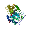
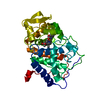
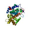
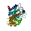
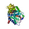
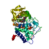
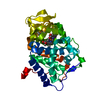
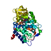
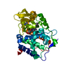
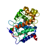
Entry Database : PDB / ID : 5ejxTitle X-ray Free Electron Laser Structure of Cytochrome C Peroxidase Cytochrome c peroxidase, mitochondrial Keywords Function / homology Function Domain/homology Component
/ / / / / / / / / / / / / / / / / / / / / / / / / / / / / / / / / / Biological species Saccharomyces cerevisiae (brewer's yeast)Method / / / Resolution : 1.5 Å Authors Doukov, T. / Soltis, S.M. / Baxter, E.L. / Cohen, A. / Song, J. / McPhillips, S. / Poulos, T.L. / Meharenna, Y.T. / Chreifi, G. Funding support Organization Grant number Country National Institutes of Health/National Institute of General Medical Sciences (NIH/NIGMS) GM57353 National Institutes of Health/National Institute of General Medical Sciences (NIH/NIGMS) P41GM103393 Department of Energy (DOE, United States) DE-AC02-76SF00515
Journal : Proc.Natl.Acad.Sci.USA / Year : 2016Title : Crystal structure of the pristine peroxidase ferryl center and its relevance to proton-coupled electron transfer.Authors : Chreifi, G. / Baxter, E.L. / Doukov, T. / Cohen, A.E. / McPhillips, S.E. / Song, J. / Meharenna, Y.T. / Soltis, S.M. / Poulos, T.L. History Deposition Nov 2, 2015 Deposition site / Processing site Revision 1.0 Jan 20, 2016 Provider / Type Revision 1.1 Feb 3, 2016 Group Revision 1.2 Feb 17, 2016 Group Revision 1.3 Sep 27, 2017 Group / Database references / Derived calculationsCategory / pdbx_audit_support / pdbx_struct_oper_listItem / _pdbx_audit_support.funding_organization / _pdbx_struct_oper_list.symmetry_operationRevision 1.4 Feb 14, 2018 Group / Category Item / _diffrn_source.pdbx_synchrotron_siteRevision 1.5 Dec 4, 2019 Group / Category / Item Revision 1.6 Sep 27, 2023 Group / Database references / Refinement descriptionCategory chem_comp_atom / chem_comp_bond ... chem_comp_atom / chem_comp_bond / database_2 / pdbx_initial_refinement_model Item / _database_2.pdbx_database_accession
Show all Show less
 Movie
Movie Controller


 Open data
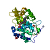
 Basic information
 Components
 Keywords
 Function and homology information
 X-RAY DIFFRACTION /
  Authors
 United States, 3items
  Citation
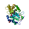
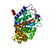
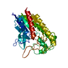
 Structure visualization
 Downloads & links
 Other downloads
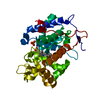


 PDBj
PDBj
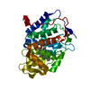
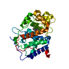
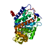
 Assembly


 Mass: 616.487 Da / Num. of mol.: 1 / Source method: obtained synthetically / Formula: C34H32FeN4O4
 Mass: 94.971 Da / Num. of mol.: 1 / Source method: obtained synthetically / Formula: PO4
 Mass: 18.015 Da / Num. of mol.: 293 / Source method: isolated from a natural source / Formula: H2O
 Sample preparation
 Processing