 Movie
Movie Controller

[English] 日本語
 Yorodumi
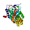
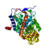
- PDB-1mkr: Crystal Structure of a Mutant Variant of Cytochrome c Peroxidase ... -
+ Open data

- Basic information
| Entry | Database: PDB / ID: 1mkr | ||||||
|---|---|---|---|---|---|---|---|
| Title | Crystal Structure of a Mutant Variant of Cytochrome c Peroxidase (Plate like crystals) | ||||||
 Components Components | Cytochrome c Peroxidase | ||||||
 Keywords Keywords | OXIDOREDUCTASE / cytochrome c peroxidase / oxygen radical / Trp cation radical / Trp-Tyr covalent cross-link | ||||||
| Function / homology |  Function and homology information Function and homology informationcytochrome-c peroxidase / cytochrome-c peroxidase activity / hydrogen peroxide catabolic process / response to reactive oxygen species / peroxidase activity / mitochondrial intermembrane space / cellular response to oxidative stress / mitochondrial matrix / heme binding / mitochondrion / metal ion binding Similarity search - Function | ||||||
| Biological species |  | ||||||
| Method |  X-RAY DIFFRACTION / MOLECULAR REPLACEMENT / Resolution: 1.58 Å X-RAY DIFFRACTION / MOLECULAR REPLACEMENT / Resolution: 1.58 Å | ||||||
 Authors Authors | Bhaskar, B. / Immoos, C.E. / Shimizu, H. / Farmer, P.J. / Poulos, T.L. | ||||||
 Citation Citation | Journal: J.Mol.Biol. / Year: 2003 Title: A Novel Heme and Peroxide-Dependent Tryptophan-Tyrosine Cross-Link in a Mutant of Cytochrome c Peroxidase Authors: Bhaskar, B. / Immoos, C.E. / Shimizu, H. / Sulc, F. / Farmer, P.J. / Poulos, T.L. | ||||||
| History |
| ||||||
| Remark 400 | COMPOUND THIS FORM OF CRYSTALS WERE PLATE LIKE, QUITE DIFFERENT FROM THE REGULAR CHUNKY FORM OF CCP. ...COMPOUND THIS FORM OF CRYSTALS WERE PLATE LIKE, QUITE DIFFERENT FROM THE REGULAR CHUNKY FORM OF CCP. THEY BELONGED TO THE SAME SPACE GROUP BUT WITH DIFFERENT UNIT CELL PARAMETERS. HELICAL BUNDLE PROTEIN SYNTHESIZED IN THE CYTOPLASM AND TRANSPORTED TO MITOCHODRIAL INTERMEMBRANE SPACE OF YEAST. IN E.COLI, HOWEVER, SYNTHESIZED AND TRANSLOCATED INTO THE PERIPLASM. PROTEIN HAS DISTINCT DISTAL AND PROXIMAL HEME POCKETS. TRP191 IN THE PROXIMAL HEME POCKET ACTS AS A CONDUIT IN ELECTRON TRANSFER WITH ITS REDOX PARTNER, CYTOCHROME C |
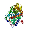
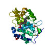
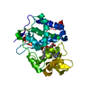
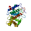
- Structure visualization
| Structure viewer | Molecule: MolmilJmol/JSmol |
|---|
- Downloads & links
-Download
| PDBx/mmCIF format | 1mkr.cif.gz | 83.2 KB | Display | PDBx/mmCIF format |
|---|---|---|---|---|
| PDB format | pdb1mkr.ent.gz | 61.5 KB | Display | PDB format |
| PDBx/mmJSON format | 1mkr.json.gz | Tree view | PDBx/mmJSON format | |
| Others |  Other downloads Other downloads |
-Validation report
| Arichive directory | https://data.pdbj.org/pub/pdb/validation_reports/mk/1mkrftp://data.pdbj.org/pub/pdb/validation_reports/mk/1mkr | HTTPS FTP |
|---|
-Related structure data
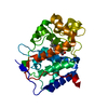
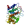
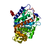
| Related structure data |  1mk8C 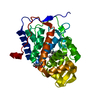 1mkqSC 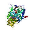 1ml2C C: citing same article ( S: Starting model for refinement |
|---|---|
| Similar structure data |


-Links
 PDBj
PDBj
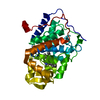
- Assembly
| Deposited unit | 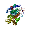
| ||||||||
|---|---|---|---|---|---|---|---|---|---|
| 1 |
| ||||||||
| Unit cell |
| ||||||||
| Details | Helical bundle protein synthesized in the cytoplasm and transported to mitochodrial intermembrane space of yeast. In E.coli, however, synthesized and translocated into the periplasm. Protein has distinct distal and proximal heme pockets. Trp191 in the proximal heme pocket acts as a conduit in electron transfer with its redox partner, cytochrome c |
-Components
| #1: Protein | Mass: 33596.266 Da / Num. of mol.: 1 / Mutation: H52Y Source method: isolated from a genetically manipulated source Source: (gene. exp.) Gene: OPBYC / Plasmid: pT7CcP / Species (production host): Escherichia coli / Production host:  |
|---|---|
| #2: Chemical | ChemComp-HEM /   Mass: 616.487 Da / Num. of mol.: 1 / Source method: obtained synthetically / Formula: C34H32FeN4O4 Mass: 616.487 Da / Num. of mol.: 1 / Source method: obtained synthetically / Formula: C34H32FeN4O4 |
| #3: Water | ChemComp-HOH /  Mass: 18.015 Da / Num. of mol.: 428 / Source method: isolated from a natural source / Formula: H2O Mass: 18.015 Da / Num. of mol.: 428 / Source method: isolated from a natural source / Formula: H2O |
-Experimental details
-Experiment
| Experiment | Method: X-RAY DIFFRACTION / Number of used crystals: 1 |
|---|
- Sample preparation
| Crystal | Density Matthews: 2.27 Å3/Da / Density % sol: 45.92 % | ||||||||||||||||||||||||||||||
|---|---|---|---|---|---|---|---|---|---|---|---|---|---|---|---|---|---|---|---|---|---|---|---|---|---|---|---|---|---|---|---|
| Crystal grow | Temperature: 277 K / Method: vapor diffusion, sitting drop / pH: 6 Details: 2-Methyl-2,4-Pentanediol (MPD), Tris-phosphate buffer, pH 6.0, VAPOR DIFFUSION, SITTING DROP, temperature 277K | ||||||||||||||||||||||||||||||
| Crystal grow | *PLUS Temperature: 4 ℃ / Method: vapor diffusion | ||||||||||||||||||||||||||||||
| Components of the solutions | *PLUS
|
-Data collection
| Diffraction | Mean temperature: 124 K |
|---|---|
| Diffraction source | Source: ROTATING ANODE / Type: RIGAKU / Wavelength: 1.54178 Å |
| Detector | Type: RIGAKU RAXIS IV / Detector: IMAGE PLATE / Date: Mar 21, 2002 / Details: Mirrors |
| Radiation | Monochromator: Graphite / Protocol: SINGLE WAVELENGTH / Monochromatic (M) / Laue (L): M / Scattering type: x-ray |
| Radiation wavelength | Wavelength: 1.54178 Å / Relative weight: 1 |
| Reflection | Resolution: 1.58→100 Å / Num. obs: 42967 / % possible obs: 94.8 % / Observed criterion σ(F): 2 / Observed criterion σ(I): 2 / Redundancy: 14.602 % / Rmerge(I) obs: 0.049 / Rsym value: 0.044 / Net I/σ(I): 37.19 |
| Reflection shell | Resolution: 1.58→1.61 Å / Redundancy: 4.24 % / Rmerge(I) obs: 0.394 / Mean I/σ(I) obs: 2.72 / Num. unique all: 1824 / Rsym value: 0.356 / % possible all: 87.9 |
| Reflection | *PLUS % possible obs: 94.8 % / Num. measured all: 627338 |
| Reflection shell | *PLUS % possible obs: 87.9 % |
- Processing
| Software |
| ||||||||||||||||||||||||||||||||||||||||||||||||||||||||||||
|---|---|---|---|---|---|---|---|---|---|---|---|---|---|---|---|---|---|---|---|---|---|---|---|---|---|---|---|---|---|---|---|---|---|---|---|---|---|---|---|---|---|---|---|---|---|---|---|---|---|---|---|---|---|---|---|---|---|---|---|---|---|
| Refinement | Method to determine structure: MOLECULAR REPLACEMENT Starting model: H52Y6 Dataset - PDB Entry - 1MKQ Resolution: 1.58→50 Å / Isotropic thermal model: Isotropic / Cross valid method: THROUGHOUT / σ(F): 0 / σ(I): 0 / Stereochemistry target values: Engh & Huber
| ||||||||||||||||||||||||||||||||||||||||||||||||||||||||||||
| Displacement parameters | Biso mean: 21.71 Å2 | ||||||||||||||||||||||||||||||||||||||||||||||||||||||||||||
| Refine analyze |
| ||||||||||||||||||||||||||||||||||||||||||||||||||||||||||||
| Refinement step | Cycle: LAST / Resolution: 1.58→50 Å
| ||||||||||||||||||||||||||||||||||||||||||||||||||||||||||||
| Refine LS restraints |
| ||||||||||||||||||||||||||||||||||||||||||||||||||||||||||||
| LS refinement shell | Refine-ID: X-RAY DIFFRACTION / Total num. of bins used: 9
| ||||||||||||||||||||||||||||||||||||||||||||||||||||||||||||
| Refinement | *PLUS % reflection Rfree: 4.8 % / Rfactor Rfree: 0.219 | ||||||||||||||||||||||||||||||||||||||||||||||||||||||||||||
| Solvent computation | *PLUS | ||||||||||||||||||||||||||||||||||||||||||||||||||||||||||||
| Displacement parameters | *PLUS | ||||||||||||||||||||||||||||||||||||||||||||||||||||||||||||
| Refine LS restraints | *PLUS
|
