 Movie
Movie Controller

[English] 日本語
 Yorodumi
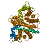
- PDB-2gb8: Solution structure of the complex between yeast iso-1-cytochrome ... -
+ Open data

- Basic information
| Entry | Database: PDB / ID: 2gb8 | ||||||
|---|---|---|---|---|---|---|---|
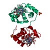
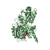
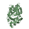
| Title | Solution structure of the complex between yeast iso-1-cytochrome c and yeast cytochrome c peroxidase | ||||||
 Components Components |
| ||||||
 Keywords Keywords | OXIDOREDUCTASE/ELECTRON TRANSPORT / protein-protein complex / electron transfer / transient complex / OXIDOREDUCTASE-ELECTRON TRANSPORT COMPLEX | ||||||
| Function / homology |  Function and homology information Function and homology informationRelease of apoptotic factors from the mitochondria / Pyroptosis / Detoxification of Reactive Oxygen Species / Respiratory electron transport / cytochrome-c peroxidase / cytochrome-c peroxidase activity / cardiolipin binding / mitochondrial electron transport, cytochrome c to oxygen / mitochondrial electron transport, ubiquinol to cytochrome c / hydrogen peroxide catabolic process ...Release of apoptotic factors from the mitochondria / Pyroptosis / Detoxification of Reactive Oxygen Species / Respiratory electron transport / cytochrome-c peroxidase / cytochrome-c peroxidase activity / cardiolipin binding / mitochondrial electron transport, cytochrome c to oxygen / mitochondrial electron transport, ubiquinol to cytochrome c / hydrogen peroxide catabolic process / response to reactive oxygen species / peroxidase activity / mitochondrial intermembrane space / cellular response to oxidative stress / electron transfer activity / mitochondrial matrix / heme binding / mitochondrion / metal ion binding Similarity search - Function | ||||||
| Biological species |  | ||||||
| Method | SOLUTION NMR / rigid-body docking solely on the basis of experimental data; sidechain dynamics. | ||||||
 Authors Authors | Volkov, A.N. / Worrall, J.A.R. / Ubbink, M. | ||||||
 Citation Citation | Journal: Proc.Natl.Acad.Sci.Usa / Year: 2006 Title: Solution structure and dynamics of the complex between cytochrome c and cytochrome c peroxidase determined by paramagnetic NMR. Authors: Volkov, A.N. / Worrall, J.A. / Holtzmann, E. / Ubbink, M. | ||||||
| History |
|
- Structure visualization
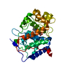
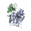
| Structure viewer | Molecule: MolmilJmol/JSmol |
|---|
- Downloads & links
-Download
| PDBx/mmCIF format | 2gb8.cif.gz | 2.6 MB | Display | PDBx/mmCIF format |
|---|---|---|---|---|
| PDB format | pdb2gb8.ent.gz | 2.3 MB | Display | PDB format |
| PDBx/mmJSON format | 2gb8.json.gz | Tree view | PDBx/mmJSON format | |
| Others |  Other downloads Other downloads |
-Validation report
| Arichive directory | https://data.pdbj.org/pub/pdb/validation_reports/gb/2gb8ftp://data.pdbj.org/pub/pdb/validation_reports/gb/2gb8 | HTTPS FTP |
|---|
-Related structure data
| Related structure data | |
|---|---|
| Similar structure data |


-Links
 PDBj
PDBj


- Assembly
| Deposited unit | 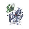
| |||||||||
|---|---|---|---|---|---|---|---|---|---|---|
| 1 |
| |||||||||
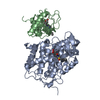
| NMR ensembles |
|
-Components
| #1: Protein | Mass: 33525.258 Da / Num. of mol.: 1 / Fragment: cytochrome c peroxidase Source method: isolated from a genetically manipulated source Source: (gene. exp.) Strain: DBY939 / Gene: CCP1 / Plasmid: CCP(MKT) / Species (production host): Escherichia coli / Production host:  |
|---|---|
| #2: Protein | Mass: 12073.835 Da / Num. of mol.: 1 Source method: isolated from a genetically manipulated source Source: (gene. exp.) Strain: Oviformis / Gene: CYC1 / Plasmid: pBTR1 / Species (production host): Escherichia coli / Production host: |
| #3: Chemical | ChemComp-HEM /   Mass: 616.487 Da / Num. of mol.: 1 / Source method: obtained synthetically / Formula: C34H32FeN4O4 Mass: 616.487 Da / Num. of mol.: 1 / Source method: obtained synthetically / Formula: C34H32FeN4O4 |
| #4: Chemical | ChemComp-HEC /   Mass: 618.503 Da / Num. of mol.: 1 / Source method: obtained synthetically / Formula: C34H34FeN4O4 Mass: 618.503 Da / Num. of mol.: 1 / Source method: obtained synthetically / Formula: C34H34FeN4O4 |
| Has protein modification | Y |
-Experimental details
-Experiment
| Experiment | Method: SOLUTION NMR |
|---|---|
| NMR experiment | Type: 1H-15N  HSQC HSQC |
| NMR details | Text: Five single-cysteine CcP variants have been prepared and labelled with a paramagnetic spin-label. For each variant, two 2D [1H,15N] HSQC spectra were acquired, one of the complex between the ...Text: Five single-cysteine CcP variants have been prepared and labelled with a paramagnetic spin-label. For each variant, two 2D [1H,15N] HSQC spectra were acquired, one of the complex between the spin-labelled protein and 15N Cc and the other of the control sample containing the complex of diamagnetically-labelled CcP with 15N Cc. From these, spin-label induced paramagnetic relaxation enhancements (PREs) of 15N Cc backbone amide resonances were determined and converted into intermolecular distance restraints, which were used for subsequent structure calculation of the protein complex. |
- Sample preparation
| Details | Contents: 0.3-0.4mM Cc(Fe3+)-CcP(Fe3+), 1:1 complex; 20mM NaPi, 100mM NaCl pH 6.0 Solvent system: 20mM NaPi, 100mM NaCl pH 6.0 |
|---|---|
| Sample conditions | Ionic strength: 120mM / pH: 6 / Pressure: ambient / Temperature: 301 K |
-NMR measurement
| NMR spectrometer | Type: Bruker DMX / Manufacturer: Bruker / Model: DMX / Field strength: 600 MHz |
|---|
- Processing
| NMR software |
| ||||||||||||||||||||||||
|---|---|---|---|---|---|---|---|---|---|---|---|---|---|---|---|---|---|---|---|---|---|---|---|---|---|
| Refinement | Method: rigid-body docking solely on the basis of experimental data; sidechain dynamics. Software ordinal: 1 Details: Coordinates of both proteins were taken from the PDB entry 2PCC. Sequences differ slightly from the experiment. Structure refinement was based on PRE-derived distance restraints for backbone ...Details: Coordinates of both proteins were taken from the PDB entry 2PCC. Sequences differ slightly from the experiment. Structure refinement was based on PRE-derived distance restraints for backbone atoms as a sole input. Only two energy terms, corresponding to restraints and van der Waals forces, are specified during the refinement procedure, which consist of two steps. First, a rigid-body docking of the protein molecules is carried out with van der Waals parameters for MTSL atoms set to zero. For each run performed, a single cluster of low-energy solutions is consistently produced. During the second step, 30 to 40 best structures are subjected to energy minimization and side-chain dynamics with fixed positions of backbone atoms for both proteins and active van der Waals parameters for MTSL. For the refined structures, the entire docking procedure is repeated until no further reduction in energy is observed. Best twenty structures of the final solution show an average rmsd from the lowest energy structure of 0.7 (0.2) Angstrom for the backbone atoms of cytochrome c after superposition of the peroxidase molecules. | ||||||||||||||||||||||||
| NMR representative | Selection criteria: lowest energy | ||||||||||||||||||||||||
| NMR ensemble | Conformer selection criteria: structures with the lowest energy Conformers calculated total number: 100 / Conformers submitted total number: 20 |
