 Movie
Movie Controller

[English] 日本語
 Yorodumi
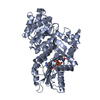
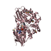
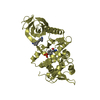
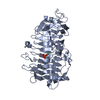
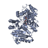
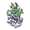
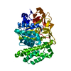
- PDB-1dkl: CRYSTAL STRUCTURE OF ESCHERICHIA COLI PHYTASE AT PH 4.5 (NO LIGAN... -
+ Open data

- Basic information
| Entry | Database: PDB / ID: 1dkl | ||||||
|---|---|---|---|---|---|---|---|
| Title | CRYSTAL STRUCTURE OF ESCHERICHIA COLI PHYTASE AT PH 4.5 (NO LIGAND BOUND) | ||||||
 Components Components | PHYTASE | ||||||
 Keywords Keywords | HYDROLASE / HISTIDINE ACID PHOSPHATASE FOLD | ||||||
| Function / homology |  Function and homology information Function and homology informationinositol phosphate phosphatase activity / cellular response to anoxia / nucleotidase activity / sugar-phosphatase activity / Hydrolases; Acting on ester bonds; Phosphoric-monoester hydrolases / cellular response to phosphate starvation / Hydrolases; Acting on acid anhydrides; In phosphorus-containing anhydrides / outer membrane-bounded periplasmic space / GTPase activity Similarity search - Function | ||||||
| Biological species |  | ||||||
| Method |  X-RAY DIFFRACTION / Resolution: 2.3 Å X-RAY DIFFRACTION / Resolution: 2.3 Å | ||||||
 Authors Authors | Lim, D. / Golovan, S. / Forsberg, C.W. / Jia, Z. | ||||||
 Citation Citation | Journal: Nat.Struct.Biol. / Year: 2000 Title: Crystal structures of Escherichia coli phytase and its complex with phytate. Authors: Lim, D. / Golovan, S. / Forsberg, C.W. / Jia, Z. #1: Journal: Acta Crystallogr.,Sect.D / Year: 1998Title: Purification, Crystallization and Preliminary X-ray Analysis of the Escherichia coli Phytase Authors: Jia, Z. / Golovan, S. / Ye, Q. / Forsberg, C.W. #2: Journal: J.Bacteriol. / Year: 1990Title: The Complete Nucleotide Sequence of the Escherichia coli Gene appA Reveals Significant Homology Between pH 2.5 Acid Phosphatase and Glucose-1-phosphatase Authors: Dassa, J. / Marck, C. / Boquet, P.L. | ||||||
| History |
|
- Structure visualization
| Structure viewer | Molecule: MolmilJmol/JSmol |
|---|
- Downloads & links
-Download
| PDBx/mmCIF format | 1dkl.cif.gz | 168.2 KB | Display | PDBx/mmCIF format |
|---|---|---|---|---|
| PDB format | pdb1dkl.ent.gz | 133.9 KB | Display | PDB format |
| PDBx/mmJSON format | 1dkl.json.gz | Tree view | PDBx/mmJSON format | |
| Others |  Other downloads Other downloads |
-Validation report
| Arichive directory | https://data.pdbj.org/pub/pdb/validation_reports/dk/1dklftp://data.pdbj.org/pub/pdb/validation_reports/dk/1dkl | HTTPS FTP |
|---|
-Related structure data


-Links
 PDBj
PDBj


- Assembly
| Deposited unit | 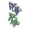
| ||||||||
|---|---|---|---|---|---|---|---|---|---|
| 1 | 
| ||||||||
| 2 | 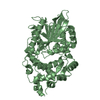
| ||||||||
| Unit cell |
| ||||||||
| Details | The biological assembly is a monomer of the dimer in the asymmetric unit. |
-Components
| #1: Protein | Mass: 44733.652 Da / Num. of mol.: 2 Source method: isolated from a genetically manipulated source Source: (gene. exp.) #2: Water | ChemComp-HOH / |  Mass: 18.015 Da / Num. of mol.: 343 / Source method: isolated from a natural source / Formula: H2O Mass: 18.015 Da / Num. of mol.: 343 / Source method: isolated from a natural source / Formula: H2OHas protein modification | Y | |
|---|
-Experimental details
-Experiment
| Experiment | Method: X-RAY DIFFRACTION / Number of used crystals: 1 |
|---|
- Sample preparation
| Crystal | Density Matthews: 2.48 Å3/Da / Density % sol: 50.45 % | ||||||||||||||||||||||||||||||||||||
|---|---|---|---|---|---|---|---|---|---|---|---|---|---|---|---|---|---|---|---|---|---|---|---|---|---|---|---|---|---|---|---|---|---|---|---|---|---|
| Crystal grow | Temperature: 273 K / Method: small tubes / pH: 4.5 Details: SODIUM ACETATE, pH 4.5, SMALL TUBES, temperature 273K | ||||||||||||||||||||||||||||||||||||
| Crystal grow | *PLUS pH: 6.6 / Method: vapor diffusion | ||||||||||||||||||||||||||||||||||||
| Components of the solutions | *PLUS
|
-Data collection
| Diffraction | Mean temperature: 277 K |
|---|---|
| Diffraction source | Source: ROTATING ANODE / Type: RIGAKU RU200 / Wavelength: 1.5418 |
| Detector | Type: MARRESEARCH / Detector: IMAGE PLATE / Date: Jul 21, 1997 |
| Radiation | Protocol: SINGLE WAVELENGTH / Monochromatic (M) / Laue (L): M / Scattering type: x-ray |
| Radiation wavelength | Wavelength: 1.5418 Å / Relative weight: 1 |
| Reflection | Resolution: 2.3→25 Å / Num. all: 106657 / Num. obs: 37076 / % possible obs: 95 % / Observed criterion σ(F): 0 / Observed criterion σ(I): 0 / Redundancy: 2.9 % / Biso Wilson estimate: 18.4 Å2 / Rmerge(I) obs: 0.085 / Net I/σ(I): 12.46 |
| Reflection shell | Resolution: 2.3→2.38 Å / Redundancy: 2.8 % / Rmerge(I) obs: 0.231 / % possible all: 97 |
| Reflection shell | *PLUS % possible obs: 97 % |
- Processing
| Software |
| ||||||||||||||||||||||||||||||||||||||||||||||||||||||||||||||||||||||||||||||||
|---|---|---|---|---|---|---|---|---|---|---|---|---|---|---|---|---|---|---|---|---|---|---|---|---|---|---|---|---|---|---|---|---|---|---|---|---|---|---|---|---|---|---|---|---|---|---|---|---|---|---|---|---|---|---|---|---|---|---|---|---|---|---|---|---|---|---|---|---|---|---|---|---|---|---|---|---|---|---|---|---|---|
| Refinement | Resolution: 2.3→25 Å / Rfactor Rfree error: 0.003 / Data cutoff high absF: 1606397.25 / Data cutoff low absF: 0 / Isotropic thermal model: RESTRAINED / Cross valid method: THROUGHOUT / σ(F): 0 / σ(I): 0 / Stereochemistry target values: ENGH & HUBER / Details: MAXIMUM LIKELIHOOD
| ||||||||||||||||||||||||||||||||||||||||||||||||||||||||||||||||||||||||||||||||
| Solvent computation | Solvent model: FLAT MODEL / Bsol: 29.82 Å2 / ksol: 0.291 e/Å3 | ||||||||||||||||||||||||||||||||||||||||||||||||||||||||||||||||||||||||||||||||
| Displacement parameters | Biso mean: 29.9 Å2
| ||||||||||||||||||||||||||||||||||||||||||||||||||||||||||||||||||||||||||||||||
| Refine analyze |
| ||||||||||||||||||||||||||||||||||||||||||||||||||||||||||||||||||||||||||||||||
| Refinement step | Cycle: LAST / Resolution: 2.3→25 Å
| ||||||||||||||||||||||||||||||||||||||||||||||||||||||||||||||||||||||||||||||||
| Refine LS restraints |
| ||||||||||||||||||||||||||||||||||||||||||||||||||||||||||||||||||||||||||||||||
| LS refinement shell | Resolution: 2.3→2.44 Å / Rfactor Rfree error: 0.01 / Total num. of bins used: 6
| ||||||||||||||||||||||||||||||||||||||||||||||||||||||||||||||||||||||||||||||||
| Xplor file |
|
