 Movie
Movie Controller

[English] 日本語
 Yorodumi
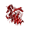
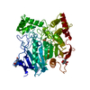
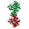
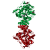
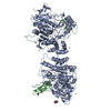
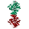
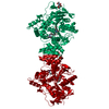
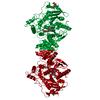
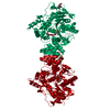
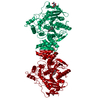
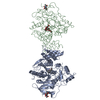
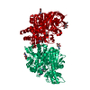
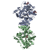
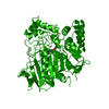
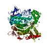
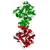
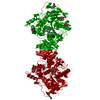
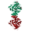
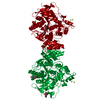
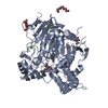
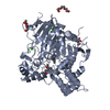
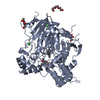
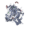
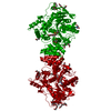
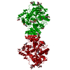
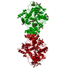
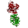
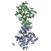
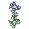
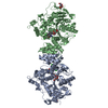
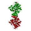
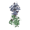
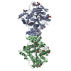
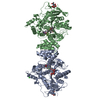
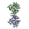
- PDB-2xi4: Torpedo californica Acetylcholinesterase in Complex with Aflatoxi... -
+ Open data

- Basic information
| Entry | Database: PDB / ID: 2xi4 | ||||||
|---|---|---|---|---|---|---|---|
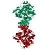
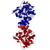
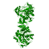
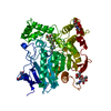
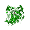
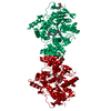
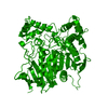
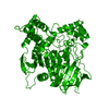
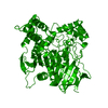
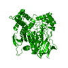
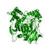
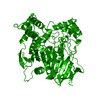
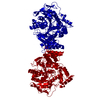
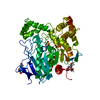
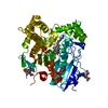
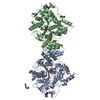
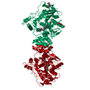
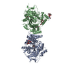
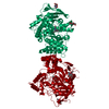
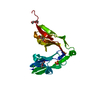
| Title | Torpedo californica Acetylcholinesterase in Complex with Aflatoxin B1 (Orthorhombic Space Group) | ||||||
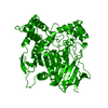
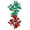
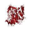
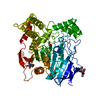
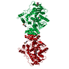
 Components Components | ACETYLCHOLINESTERASE | ||||||
 Keywords Keywords | HYDROLASE / ACETYLCHOLINESTERASE BACKDOOR / MYCOTOXIN / ALZHEIMER DISEASE / CELL JUNCTION / GPI-ANCHOR / NEUROTRANSMITTER CLEAVAGE / NEUROTRANSMITTER DEGRADATION / SERINE ESTERASE / SYNAPSE | ||||||
| Function / homology |  Function and homology information Function and homology informationacetylcholine catabolic process in synaptic cleft / acetylcholinesterase / choline metabolic process / acetylcholinesterase activity / side of membrane / synaptic cleft / synapse / : / plasma membrane Similarity search - Function | ||||||
| Biological species |   TORPEDO CALIFORNICA (Pacific electric ray) TORPEDO CALIFORNICA (Pacific electric ray) | ||||||
| Method |  X-RAY DIFFRACTION / SYNCHROTRON / MOLECULAR REPLACEMENT / Resolution: 2.3 Å X-RAY DIFFRACTION / SYNCHROTRON / MOLECULAR REPLACEMENT / Resolution: 2.3 Å | ||||||
 Authors Authors | Sanson, B. / Colletier, J.P. / Xu, Y. / Lang, P.T. / Jiang, H. / Silman, I. / Sussman, J.L. / Weik, M. | ||||||
 Citation Citation | Journal: Protein Sci. / Year: 2011 Title: Backdoor Opening Mechanism in Acetylcholinesterase Based on X-Ray Crystallography and Molecular Dynamics Simulations. Authors: Sanson, B. / Colletier, J.P. / Xu, Y. / Lang, P.T. / Jiang, H. / Silman, I. / Sussman, J.L. / Weik, M. | ||||||
| History |
|
- Structure visualization
| Structure viewer | Molecule: MolmilJmol/JSmol |
|---|
- Downloads & links
-Download
| PDBx/mmCIF format | 2xi4.cif.gz | 451.1 KB | Display | PDBx/mmCIF format |
|---|---|---|---|---|
| PDB format | pdb2xi4.ent.gz | 375.2 KB | Display | PDB format |
| PDBx/mmJSON format | 2xi4.json.gz | Tree view | PDBx/mmJSON format | |
| Others |  Other downloads Other downloads |
-Validation report
| Arichive directory | https://data.pdbj.org/pub/pdb/validation_reports/xi/2xi4ftp://data.pdbj.org/pub/pdb/validation_reports/xi/2xi4 | HTTPS FTP |
|---|
-Related structure data
| Related structure data | 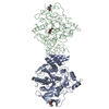 1w75S S: Starting model for refinement |
|---|---|
| Similar structure data |


-Links
 PDBj
PDBj
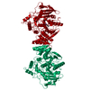
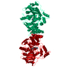
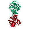
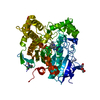
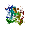
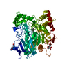
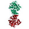
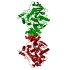
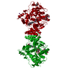
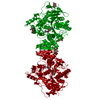
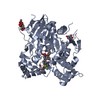
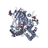
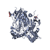
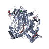
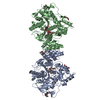
- Assembly
| Deposited unit | 
| ||||||||
|---|---|---|---|---|---|---|---|---|---|
| 1 |
| ||||||||
| Unit cell |
|
-Components
-Protein / Sugars , 2 types, 7 molecules AB
| #1: Protein | Mass: 60736.516 Da / Num. of mol.: 2 / Fragment: RESIDUES 22-558 / Source method: isolated from a natural source Source: (natural) TORPEDO CALIFORNICA (Pacific electric ray)Organ: ELECTRIC ORGAN / Variant: G2 FORM / Tissue: ELECTROPLAQUE / References: UniProt: P04058, acetylcholinesterase #6: Sugar | ChemComp-NAG /  Type: D-saccharide, beta linking / Mass: 221.208 Da / Num. of mol.: 5 Type: D-saccharide, beta linking / Mass: 221.208 Da / Num. of mol.: 5Source method: isolated from a genetically manipulated source Formula: C8H15NO6 |
|---|
-Non-polymers , 6 types, 759 molecules 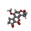


| #2: Chemical |  Mass: 312.274 Da / Num. of mol.: 2 / Source method: obtained synthetically / Formula: C17H12O6 / Comment: toxin*YM Mass: 312.274 Da / Num. of mol.: 2 / Source method: obtained synthetically / Formula: C17H12O6 / Comment: toxin*YM#3: Chemical | ChemComp-CL /  Mass: 35.453 Da / Num. of mol.: 4 / Source method: obtained synthetically / Formula: Cl Mass: 35.453 Da / Num. of mol.: 4 / Source method: obtained synthetically / Formula: Cl#4: Chemical | ChemComp-PEG /  Mass: 106.120 Da / Num. of mol.: 6 / Source method: obtained synthetically / Formula: C4H10O3 Mass: 106.120 Da / Num. of mol.: 6 / Source method: obtained synthetically / Formula: C4H10O3#5: Chemical |  Mass: 150.173 Da / Num. of mol.: 2 / Source method: obtained synthetically / Formula: C6H14O4 Mass: 150.173 Da / Num. of mol.: 2 / Source method: obtained synthetically / Formula: C6H14O4#7: Chemical | ChemComp-K / |  Mass: 39.098 Da / Num. of mol.: 1 / Source method: obtained synthetically / Formula: K Mass: 39.098 Da / Num. of mol.: 1 / Source method: obtained synthetically / Formula: K#8: Water | ChemComp-HOH / | Mass: 18.015 Da / Num. of mol.: 744 / Source method: isolated from a natural source / Formula: H2O |
|---|
-Details
| Has protein modification | Y |
|---|
-Experimental details
-Experiment
| Experiment | Method: X-RAY DIFFRACTION / Number of used crystals: 1 |
|---|
- Sample preparation
| Crystal | Density Matthews: 3.1 Å3/Da / Density % sol: 60.2 % / Description: NONE |
|---|---|
| Crystal grow | Temperature: 277 K / pH: 5.8 Details: ACHE WAS CRYSTALLIZED IN 34% PEG200, 150 MM MES, PH 6, 4 DEG. C. THE CRYSTAL WAS SOAKED FOR 15 H IN 100 MICROMOLAR AFLATOXIN B1, 10% ETOH, 36% PEG200, 150MM MES, PH 5.8, 4 DEG C. |
-Data collection
| Diffraction | Mean temperature: 100 K |
|---|---|
| Diffraction source | Source: SYNCHROTRON / Site: ESRF  / Beamline: ID14-2 / Wavelength: 0.933 / Beamline: ID14-2 / Wavelength: 0.933 |
| Detector | Type: ADSC CCD / Detector: CCD / Date: Sep 10, 2007 |
| Radiation | Protocol: SINGLE WAVELENGTH / Monochromatic (M) / Laue (L): M / Scattering type: x-ray |
| Radiation wavelength | Wavelength: 0.933 Å / Relative weight: 1 |
| Reflection | Resolution: 2.3→46.22 Å / Num. obs: 65334 / % possible obs: 98.1 % / Observed criterion σ(I): -3 / Redundancy: 6.05 % / Biso Wilson estimate: 38.486 Å2 / Rmerge(I) obs: 0.06 / Net I/σ(I): 23.07 |
| Reflection shell | Resolution: 2.3→2.35 Å / Redundancy: 5.94 % / Rmerge(I) obs: 0.42 / Mean I/σ(I) obs: 4.57 / % possible all: 100 |
- Processing
| Software |
| ||||||||||||||||||||||||||||||||||||||||||||||||||||||||||||||||||||||||||||||||||||||||||||||||||||||||||||||||||||||||||||||||||||||||||||||||||||||||||||||||||||||||||||||||||||||
|---|---|---|---|---|---|---|---|---|---|---|---|---|---|---|---|---|---|---|---|---|---|---|---|---|---|---|---|---|---|---|---|---|---|---|---|---|---|---|---|---|---|---|---|---|---|---|---|---|---|---|---|---|---|---|---|---|---|---|---|---|---|---|---|---|---|---|---|---|---|---|---|---|---|---|---|---|---|---|---|---|---|---|---|---|---|---|---|---|---|---|---|---|---|---|---|---|---|---|---|---|---|---|---|---|---|---|---|---|---|---|---|---|---|---|---|---|---|---|---|---|---|---|---|---|---|---|---|---|---|---|---|---|---|---|---|---|---|---|---|---|---|---|---|---|---|---|---|---|---|---|---|---|---|---|---|---|---|---|---|---|---|---|---|---|---|---|---|---|---|---|---|---|---|---|---|---|---|---|---|---|---|---|---|
| Refinement | Method to determine structure: MOLECULAR REPLACEMENT Starting model: PDB ENTRY 1W75 Resolution: 2.3→46.22 Å / Cor.coef. Fo:Fc: 0.959 / Cor.coef. Fo:Fc free: 0.932 / SU B: 13.367 / SU ML: 0.148 / Cross valid method: THROUGHOUT / ESU R: 0.264 / ESU R Free: 0.214 / Stereochemistry target values: MAXIMUM LIKELIHOOD / Details: HYDROGENS HAVE BEEN ADDED IN THE RIDING POSITIONS.
| ||||||||||||||||||||||||||||||||||||||||||||||||||||||||||||||||||||||||||||||||||||||||||||||||||||||||||||||||||||||||||||||||||||||||||||||||||||||||||||||||||||||||||||||||||||||
| Solvent computation | Ion probe radii: 0.8 Å / Shrinkage radii: 0.8 Å / VDW probe radii: 1.4 Å / Solvent model: BABINET MODEL WITH MASK | ||||||||||||||||||||||||||||||||||||||||||||||||||||||||||||||||||||||||||||||||||||||||||||||||||||||||||||||||||||||||||||||||||||||||||||||||||||||||||||||||||||||||||||||||||||||
| Displacement parameters | Biso mean: 37.284 Å2
| ||||||||||||||||||||||||||||||||||||||||||||||||||||||||||||||||||||||||||||||||||||||||||||||||||||||||||||||||||||||||||||||||||||||||||||||||||||||||||||||||||||||||||||||||||||||
| Refinement step | Cycle: LAST / Resolution: 2.3→46.22 Å
| ||||||||||||||||||||||||||||||||||||||||||||||||||||||||||||||||||||||||||||||||||||||||||||||||||||||||||||||||||||||||||||||||||||||||||||||||||||||||||||||||||||||||||||||||||||||
| Refine LS restraints |
|