 Movie
Movie Controller

[English] 日本語
 Yorodumi
- PDB-2dfp: X-RAY STRUCTURE OF AGED DI-ISOPROPYL-PHOSPHORO-FLUORIDATE (DFP) B... -
+ Open data

- Basic information
| Entry | Database: PDB / ID: 2dfp | |||||||||
|---|---|---|---|---|---|---|---|---|---|---|
| Title | X-RAY STRUCTURE OF AGED DI-ISOPROPYL-PHOSPHORO-FLUORIDATE (DFP) BOUND TO ACETYLCHOLINESTERASE | |||||||||
 Components Components | PROTEIN (ACETYLCHOLINESTERASE) | |||||||||
 Keywords Keywords | HYDROLASE / AGED ORGANOPHOSPHATE / DFP / SERINE HYDROLASE / NEUROTRANSMITTER CLEAVAGE / CATALYTIC TRIAD / ALPHA/BETA HYDROLASE | |||||||||
| Function / homology |  Function and homology information Function and homology informationacetylcholine catabolic process in synaptic cleft / acetylcholinesterase / choline metabolic process / acetylcholinesterase activity / side of membrane / synaptic cleft / synapse / : / plasma membrane Similarity search - Function | |||||||||
| Biological species |   Torpedo californica (Pacific electric ray) Torpedo californica (Pacific electric ray) | |||||||||
| Method |  X-RAY DIFFRACTION / MOLECULAR REPLACEMENT / Resolution: 2.3 Å X-RAY DIFFRACTION / MOLECULAR REPLACEMENT / Resolution: 2.3 Å | |||||||||
 Authors Authors | Kryger, G. / Millard, C.B. / Silman, I. / Sussman, J.L. | |||||||||
 Citation Citation | Journal: Biochemistry / Year: 1999 Title: Crystal structures of aged phosphonylated acetylcholinesterase: nerve agent reaction products at the atomic level. Authors: Millard, C.B. / Kryger, G. / Ordentlich, A. / Greenblatt, H.M. / Harel, M. / Raves, M.L. / Segall, Y. / Barak, D. / Shafferman, A. / Silman, I. / Sussman, J.L. #1: Journal: Science / Year: 1991Title: Atomic Structure of Acetylcholinesterase from Torpedo Californica: A Prototypic Acetylcholine-Binding Protein Authors: Sussman, J.L. / Harel, M. / Frolow, F. / Oefner, C. / Goldman, A. / Toker, L. / Silman, I. | |||||||||
| History |
|
- Structure visualization
| Structure viewer | Molecule: MolmilJmol/JSmol |
|---|
- Downloads & links
-Download
| PDBx/mmCIF format | 2dfp.cif.gz | 132.3 KB | Display | PDBx/mmCIF format |
|---|---|---|---|---|
| PDB format | pdb2dfp.ent.gz | 100.9 KB | Display | PDB format |
| PDBx/mmJSON format | 2dfp.json.gz | Tree view | PDBx/mmJSON format | |
| Others |  Other downloads Other downloads |
-Validation report
| Arichive directory | https://data.pdbj.org/pub/pdb/validation_reports/df/2dfpftp://data.pdbj.org/pub/pdb/validation_reports/df/2dfp | HTTPS FTP |
|---|
-Related structure data
| Related structure data | 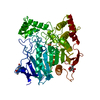 1cfjC 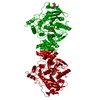 1somC 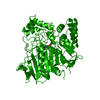 2aceS S: Starting model for refinement C: citing same article ( |
|---|---|
| Similar structure data |


-Links
 PDBj
PDBj
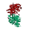
- Assembly
| Deposited unit | 
| ||||||||||
|---|---|---|---|---|---|---|---|---|---|---|---|
| 1 | 
| ||||||||||
| Unit cell |
| ||||||||||
| Components on special symmetry positions |
| ||||||||||
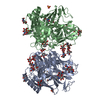
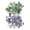
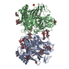
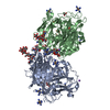
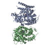
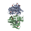
| Details | TORPEDO CALIFONICA ACETYLCHOLINESTERASE IS A G2 DIMER IN SOLUTION (SEE SUSSMAN 1988). THE ASYMMETRIC UNIT CONTAINS A MONOMER, WITH THE CRYSTALLOGRAPHIC TWO-FOLD AXIS RELATING THE TWO MONOMERS IN A DIMER. BIOMOLECULE: THE ENZYME IS A GPI-ANCHORED DIMER, THE TWO MONOMERS IN THE DIMER ARE RELATED BY THE CRYSTALLOGRAPHIC TWO-FOLD SYMMETRY AND THUS GENERATE A FOUR HELIX BUNDLE A365-A375 AND A518-A535. DFP WAS REACTED WITH THE ENZYME IN SOLUTION. THE SOLUTION OF THE INHIBITED ENZYME WAS DIALYZED TO REMOVE EXCESS ORGANOPHOSPHATE, CHECKED FOR ACTIVITY AND FOUND TO REACT LIKE "AGED" PHOSPHYLATED ACHE, AND THEN SUBJECTED TO CRYSTALLIZATION. THIS STRUCTURE IS MORE COMPLETE THAN THE STARTING MODEL OF THE NATIVE STRUCTURE (PDB ID 2ACE). RESIDUES THAT ARE NOT SEEN IN THE CRYSTAL STRUCTURE DUE TO DISORDER INCLUDE THE N-TERMINAL RESIDUE ASP 1 AND THE C-TERMINAL RESIDUES AFTER THR 535. THR 535 IS THE LAST RESIDUE OBSERVED AT THE C-TERMINUS. |
-Components
| #1: Protein | Mass: 60569.266 Da / Num. of mol.: 1 / Source method: isolated from a natural source Details: THE PROTEIN, ACETYLCHOLINESTERASE, WAS TREATED WITH THE ORGANOPHOSPHATE, DFP, PRIOR TO CRYSTALLIZATION. Source: (natural) Torpedo californica (Pacific electric ray)Organ: ELECTRIC ORGAN / Variant: G2 FORM / Tissue: ELECTROPLAQUE Keywords: THE PROTEIN, ACETYLCHOLINESTERASE, WAS TREATED WITH THE ORGANOPHOSPHATE, DFP, PRIOR TO CRYSTALLIZATIONReferences: UniProt: P04058, acetylcholinesterase | ||||||||
|---|---|---|---|---|---|---|---|---|---|
| #2: Polysaccharide | | #3: Sugar | ChemComp-NAG / |   Type: D-saccharide, beta linking / Mass: 221.208 Da / Num. of mol.: 1 / Source method: isolated from a natural source / Formula: C8H15NO6 Type: D-saccharide, beta linking / Mass: 221.208 Da / Num. of mol.: 1 / Source method: isolated from a natural source / Formula: C8H15NO6#4: Chemical | ChemComp-MES / |   Mass: 195.237 Da / Num. of mol.: 1 / Source method: obtained synthetically / Formula: C6H13NO4S / Comment: pH buffer*YM Mass: 195.237 Da / Num. of mol.: 1 / Source method: obtained synthetically / Formula: C6H13NO4S / Comment: pH buffer*YM#5: Water | ChemComp-HOH / |  Mass: 18.015 Da / Num. of mol.: 376 / Source method: isolated from a natural source / Formula: H2O Mass: 18.015 Da / Num. of mol.: 376 / Source method: isolated from a natural source / Formula: H2ONonpolymer details | DFP, AGED ORGANOPHOSPHATE MES, USED AS THE CRYSTALLIZATION BUFFER THERE ARE 5 NAG GROUPS NUMBERED ...DFP, AGED ORGANOPHOS | |
-Experimental details
-Experiment
| Experiment | Method: X-RAY DIFFRACTION / Number of used crystals: 1 |
|---|
- Sample preparation
| Crystal | Density Matthews: 3.8 Å3/Da / Density % sol: 68 % / Description: USED COMPOSITE OMIT MAP METHOD | ||||||||||||||||||||||||||||||
|---|---|---|---|---|---|---|---|---|---|---|---|---|---|---|---|---|---|---|---|---|---|---|---|---|---|---|---|---|---|---|---|
| Crystal grow | Temperature: 277 K / pH: 5.8 / Details: pH 5.8, temperature 277K | ||||||||||||||||||||||||||||||
| Components of the solutions |
| ||||||||||||||||||||||||||||||
| Crystal grow | *PLUS Temperature: 4 ℃ / Method: vapor diffusion, hanging drop / PH range low: 6 / PH range high: 5.8 | ||||||||||||||||||||||||||||||
| Components of the solutions | *PLUS
|
-Data collection
| Diffraction | Mean temperature: 100 K |
|---|---|
| Diffraction source | Source: ROTATING ANODE / Type: RIGAKU RU300 / Wavelength: 1.5418 |
| Detector | Type: RIGAKU RAXIS II / Detector: IMAGE PLATE / Date: May 15, 1998 / Details: MIRRORS |
| Radiation | Monochromator: NI FILTER / Protocol: SINGLE WAVELENGTH / Monochromatic (M) / Laue (L): M / Scattering type: x-ray |
| Radiation wavelength | Wavelength: 1.5418 Å / Relative weight: 1 |
| Reflection | Resolution: 2.3→20 Å / Num. all: 42208 / Num. obs: 42208 / % possible obs: 93.7 % / Observed criterion σ(I): 0 / Redundancy: 2.5 % / Biso Wilson estimate: 45.7 Å2 / Rsym value: 0.05 |
| Reflection shell | Resolution: 2.3→2.38 Å / % possible all: 65.2 |
| Reflection | *PLUS Num. obs: 42410 / % possible obs: 94.2 % / Num. measured all: 278106 / Rmerge(I) obs: 0.07 |
| Reflection shell | *PLUS % possible obs: 66.3 % / Rmerge(I) obs: 0.41 |
- Processing
| Software |
| ||||||||||||||||||||||||||||||||||||||||||||||||||||||||||||
|---|---|---|---|---|---|---|---|---|---|---|---|---|---|---|---|---|---|---|---|---|---|---|---|---|---|---|---|---|---|---|---|---|---|---|---|---|---|---|---|---|---|---|---|---|---|---|---|---|---|---|---|---|---|---|---|---|---|---|---|---|---|
| Refinement | Method to determine structure: MOLECULAR REPLACEMENT Starting model: PDB ENTRY 2ACE Resolution: 2.3→20 Å / Rfactor Rfree error: 0.005 / Data cutoff high rms absF: 2409412.92 / Isotropic thermal model: OVERALL / Cross valid method: THROUGHOUT / σ(F): 0 / Details: BULK SOLVENT MODEL USED
| ||||||||||||||||||||||||||||||||||||||||||||||||||||||||||||
| Solvent computation | Solvent model: FLAT MODEL / Bsol: 44.45 Å2 / ksol: 0.345 e/Å3 | ||||||||||||||||||||||||||||||||||||||||||||||||||||||||||||
| Displacement parameters | Biso mean: 42.4 Å2
| ||||||||||||||||||||||||||||||||||||||||||||||||||||||||||||
| Refine analyze |
| ||||||||||||||||||||||||||||||||||||||||||||||||||||||||||||
| Refinement step | Cycle: LAST / Resolution: 2.3→20 Å
| ||||||||||||||||||||||||||||||||||||||||||||||||||||||||||||
| Refine LS restraints |
| ||||||||||||||||||||||||||||||||||||||||||||||||||||||||||||
| LS refinement shell | Resolution: 2.3→2.38 Å / Rfactor Rfree error: 0.032 / Total num. of bins used: 10
| ||||||||||||||||||||||||||||||||||||||||||||||||||||||||||||
| Xplor file |
| ||||||||||||||||||||||||||||||||||||||||||||||||||||||||||||
| Software | *PLUS Name: CNS / Version: 0.4 / Classification: refinement | ||||||||||||||||||||||||||||||||||||||||||||||||||||||||||||
| Refinement | *PLUS Num. reflection obs: 40334 / σ(F): 0 / % reflection Rfree: 4.2 % / Rfactor obs: 0.186 | ||||||||||||||||||||||||||||||||||||||||||||||||||||||||||||
| Solvent computation | *PLUS | ||||||||||||||||||||||||||||||||||||||||||||||||||||||||||||
| Displacement parameters | *PLUS Biso mean: 42.4 Å2 | ||||||||||||||||||||||||||||||||||||||||||||||||||||||||||||
| Refine LS restraints | *PLUS
| ||||||||||||||||||||||||||||||||||||||||||||||||||||||||||||
| LS refinement shell | *PLUS Rfactor Rfree: 0.363 / % reflection Rfree: 4.5 % / Rfactor Rwork: 0.353 |
