 Movie
Movie Controller

[English] 日本語
 Yorodumi
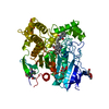
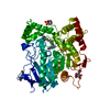
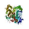
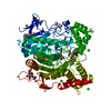
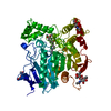
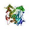
- PDB-1cfj: METHYLPHOSPHONYLATED ACETYLCHOLINESTERASE (AGED) OBTAINED BY REAC... -
+ Open data

- Basic information
| Entry | Database: PDB / ID: 1cfj | |||||||||
|---|---|---|---|---|---|---|---|---|---|---|
| Title | METHYLPHOSPHONYLATED ACETYLCHOLINESTERASE (AGED) OBTAINED BY REACTION WITH O-ISOPROPYLMETHYLPHOSPHONOFLUORIDATE (GB, SARIN) | |||||||||
 Components Components | PROTEIN (ACETYLCHOLINESTERASE) | |||||||||
 Keywords Keywords | HYDROLASE / CHOLINESTERASE / ORGANOPHOSPHATE / SERINE HYDROLASE / CHEMICAL-WARFARE | |||||||||
| Function / homology |  Function and homology information Function and homology informationacetylcholine catabolic process in synaptic cleft / acetylcholinesterase / choline metabolic process / acetylcholinesterase activity / side of membrane / synaptic cleft / synapse / : / plasma membrane Similarity search - Function | |||||||||
| Biological species |   Torpedo californica (Pacific electric ray) Torpedo californica (Pacific electric ray) | |||||||||
| Method |  X-RAY DIFFRACTION / SYNCHROTRON / MOLECULAR REPLACEMENT / Resolution: 2.6 Å X-RAY DIFFRACTION / SYNCHROTRON / MOLECULAR REPLACEMENT / Resolution: 2.6 Å | |||||||||
 Authors Authors | Millard, C.B. / Silman, I. / Sussman, J.L. | |||||||||
 Citation Citation | Journal: Biochemistry / Year: 1999 Title: Crystal structures of aged phosphonylated acetylcholinesterase: nerve agent reaction products at the atomic level. Authors: Millard, C.B. / Kryger, G. / Ordentlich, A. / Greenblatt, H.M. / Harel, M. / Raves, M.L. / Segall, Y. / Barak, D. / Shafferman, A. / Silman, I. / Sussman, J.L. #1: Journal: Structure and Function of Cholinesterases and Related ProteinsYear: 1998 Title: Crystal Structural of "Aged" Phosphorylated and Phosphonylated torpedo californica Acetylcholinesterase Authors: Millard, C.B. / Kryger, G. / Ordentlich, A. / Harel, M. / Raves, M.L. / Greenblatt, H.M. / Segall, Y. / Barak, D. / Shafferman, A. / Silman, I. / Sussman, J.L. | |||||||||
| History |
|
- Structure visualization
| Structure viewer | Molecule: MolmilJmol/JSmol |
|---|
- Downloads & links
-Download
| PDBx/mmCIF format | 1cfj.cif.gz | 123.5 KB | Display | PDBx/mmCIF format |
|---|---|---|---|---|
| PDB format | pdb1cfj.ent.gz | 95.3 KB | Display | PDB format |
| PDBx/mmJSON format | 1cfj.json.gz | Tree view | PDBx/mmJSON format | |
| Others |  Other downloads Other downloads |
-Validation report
| Arichive directory | https://data.pdbj.org/pub/pdb/validation_reports/cf/1cfjftp://data.pdbj.org/pub/pdb/validation_reports/cf/1cfj | HTTPS FTP |
|---|
-Related structure data
| Related structure data | 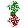 1somC 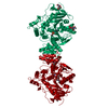 2dfpC 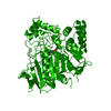 2aceS S: Starting model for refinement C: citing same article ( |
|---|---|
| Similar structure data |


-Links
 PDBj
PDBj
- Assembly
| Deposited unit | 
| ||||||||
|---|---|---|---|---|---|---|---|---|---|
| 1 |
| ||||||||
| Unit cell |
|
-Components
| #1: Protein | Mass: 60736.516 Da / Num. of mol.: 1 / Source method: isolated from a natural source Source: (natural) Torpedo californica (Pacific electric ray)Cellular location: MEMBRANE BOUND BY GPI ANCHOR / Organ: ELECTRIC ORGAN / Tissue: ELECTROPLAQUE / References: UniProt: P04058, acetylcholinesterase | ||||||||
|---|---|---|---|---|---|---|---|---|---|
| #2: Sugar |   Type: D-saccharide, beta linking / Mass: 221.208 Da / Num. of mol.: 2 Type: D-saccharide, beta linking / Mass: 221.208 Da / Num. of mol.: 2Source method: isolated from a genetically manipulated source Formula: C8H15NO6 #3: Chemical | ChemComp-GB / | 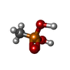  Mass: 96.022 Da / Num. of mol.: 1 / Source method: obtained synthetically / Formula: CH5O3P Mass: 96.022 Da / Num. of mol.: 1 / Source method: obtained synthetically / Formula: CH5O3P#4: Water | ChemComp-HOH / |  Mass: 18.015 Da / Num. of mol.: 220 / Source method: isolated from a natural source / Formula: H2O Mass: 18.015 Da / Num. of mol.: 220 / Source method: isolated from a natural source / Formula: H2OHas protein modification | Y | Nonpolymer details | ANIONIC METHYLPHOS | |
-Experimental details
-Experiment
| Experiment | Method: X-RAY DIFFRACTION / Number of used crystals: 1 |
|---|
- Sample preparation
| Crystal | Density Matthews: 3.8 Å3/Da / Density % sol: 68 % | ||||||||||||||||||||||||||||||
|---|---|---|---|---|---|---|---|---|---|---|---|---|---|---|---|---|---|---|---|---|---|---|---|---|---|---|---|---|---|---|---|
| Crystal grow | Temperature: 277 K / pH: 6 Details: 35-40% (W/V) PEG-200 0.15 M MES BUFFER PH 6, 0.05 M NACL, 4 DEG C , temperature 277.0K | ||||||||||||||||||||||||||||||
| Crystal grow | *PLUS Temperature: 4 ℃ / Method: vapor diffusion, hanging drop / PH range low: 6 / PH range high: 5.8 | ||||||||||||||||||||||||||||||
| Components of the solutions | *PLUS
|
-Data collection
| Diffraction | Mean temperature: 100 K |
|---|---|
| Diffraction source | Source: SYNCHROTRON / Site: NSLS  / Beamline: X12C / Wavelength: 1.1 / Beamline: X12C / Wavelength: 1.1 |
| Detector | Type: BRANDEIS - B4 / Detector: CCD / Date: Jul 1, 1997 |
| Radiation | Protocol: SINGLE WAVELENGTH / Monochromatic (M) / Laue (L): M / Scattering type: x-ray |
| Radiation wavelength | Wavelength: 1.1 Å / Relative weight: 1 |
| Reflection | Resolution: 2.6→30 Å / Num. obs: 30011 / % possible obs: 97.4 % / Redundancy: 6.7 % / Biso Wilson estimate: 40.2 Å2 / Rsym value: 0.09 / Net I/σ(I): 10.3 |
| Reflection shell | Resolution: 2.6→2.69 Å / Mean I/σ(I) obs: 2.2 / Rsym value: 0.17 / % possible all: 99.3 |
| Reflection | *PLUS Num. measured all: 200755 / Rmerge(I) obs: 0.09 |
| Reflection shell | *PLUS % possible obs: 99.3 % / Rmerge(I) obs: 0.17 |
- Processing
| Software |
| ||||||||||||||||||||||||||||||||||||||||||||||||||||||||||||||||||||||||||||||||
|---|---|---|---|---|---|---|---|---|---|---|---|---|---|---|---|---|---|---|---|---|---|---|---|---|---|---|---|---|---|---|---|---|---|---|---|---|---|---|---|---|---|---|---|---|---|---|---|---|---|---|---|---|---|---|---|---|---|---|---|---|---|---|---|---|---|---|---|---|---|---|---|---|---|---|---|---|---|---|---|---|---|
| Refinement | Method to determine structure: MOLECULAR REPLACEMENT Starting model: 2ACE Resolution: 2.6→30 Å / Rfactor Rfree error: 0.006 / Data cutoff high rms absF: 3132443.55 / Isotropic thermal model: RESTRAINED / Cross valid method: THROUGHOUT / σ(F): 0 Details: CONTINUOUS POSITIVE DIFFERENCE DENSITY IN (FO-FC) MAPS OCCURS IN FRONT OF THE INDOLE RINGS OF W84 AND W279. THIS DENSITY IS PRESENTLY MODELLED WITH WATERS 1007/1008/1009 (W84) AND WATERS 1003/1004/1005 (W279)
| ||||||||||||||||||||||||||||||||||||||||||||||||||||||||||||||||||||||||||||||||
| Solvent computation | Solvent model: FLAT MODEL / Bsol: 38.55 Å2 / ksol: 0.346 e/Å3 | ||||||||||||||||||||||||||||||||||||||||||||||||||||||||||||||||||||||||||||||||
| Displacement parameters | Biso mean: 32.5 Å2
| ||||||||||||||||||||||||||||||||||||||||||||||||||||||||||||||||||||||||||||||||
| Refine analyze |
| ||||||||||||||||||||||||||||||||||||||||||||||||||||||||||||||||||||||||||||||||
| Refinement step | Cycle: LAST / Resolution: 2.6→30 Å
| ||||||||||||||||||||||||||||||||||||||||||||||||||||||||||||||||||||||||||||||||
| Refine LS restraints |
| ||||||||||||||||||||||||||||||||||||||||||||||||||||||||||||||||||||||||||||||||
| LS refinement shell | Resolution: 2.6→2.76 Å / Rfactor Rfree error: 0.017 / Total num. of bins used: 6
| ||||||||||||||||||||||||||||||||||||||||||||||||||||||||||||||||||||||||||||||||
| Xplor file |
| ||||||||||||||||||||||||||||||||||||||||||||||||||||||||||||||||||||||||||||||||
| Software | *PLUS Name: CNS / Version: 0.5 / Classification: refinement | ||||||||||||||||||||||||||||||||||||||||||||||||||||||||||||||||||||||||||||||||
| Refinement | *PLUS Num. reflection obs: 27034 | ||||||||||||||||||||||||||||||||||||||||||||||||||||||||||||||||||||||||||||||||
| Solvent computation | *PLUS | ||||||||||||||||||||||||||||||||||||||||||||||||||||||||||||||||||||||||||||||||
| Displacement parameters | *PLUS | ||||||||||||||||||||||||||||||||||||||||||||||||||||||||||||||||||||||||||||||||
| Refine LS restraints | *PLUS
|
