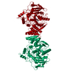
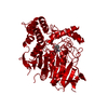
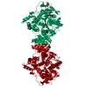
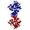
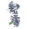
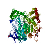
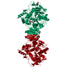
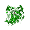
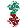
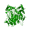
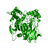
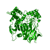
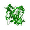
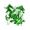
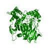
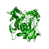
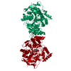
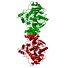
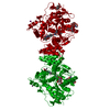
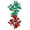
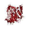
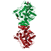
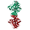
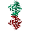
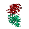
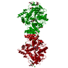
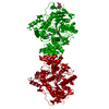
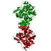
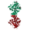
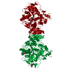
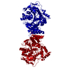
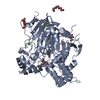
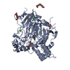
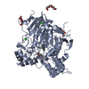
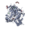
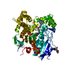
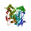
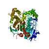
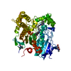
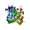
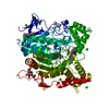
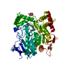
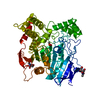
Mass: 61325.090 Da / Num. of mol.: 1 / Fragment: RESIDUES 22-464 / Source method: isolated from a natural source Details: 2-FORMYL-1-METHYL-PYRIDINIUM CHLORIDE (PRALYDOXIME, 2-PAM) IN ACTIVE SITE GORGE Source: (natural) TORPEDO CALIFORNICA (Pacific electric ray) References: UniProt: P04058, acetylcholinesterase
Resolution: 2.71→15.66 Å / Cor.coef. Fo:Fc: 0.941 / Cor.coef. Fo:Fc free: 0.886 / SU B: 10.334 / SU ML: 0.197 / Cross valid method: THROUGHOUT / ESU R: 0.359 / ESU R Free: 0.27 / Stereochemistry target values: MAXIMUM LIKELIHOOD / Details: HYDROGENS HAVE BEEN ADDED IN THE RIDING POSITIONS.
Rfactor
Num. reflection
% reflection
Selection details
Rfree
0.234
1340
5.1 %
RANDOM
Rwork
0.171
-
-
-
obs
0.174
25095
100 %
-
Solvent computation
Ion probe radii: 0.8 Å / Shrinkage radii: 0.8 Å / VDW probe radii: 1.2 Å / Solvent model: MASK
 Movie
Movie Controller


 Open data
 Basic information
 Components
 Keywords
 Function and homology information
 TORPEDO CALIFORNICA (Pacific electric ray)
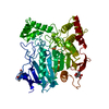
 X-RAY DIFFRACTION /
  Authors
 Citation
 Structure visualization
 Downloads & links
 Other downloads
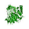


 PDBj
PDBj
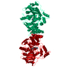
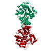
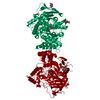
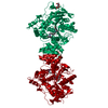
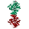
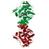
 Assembly

 Mass: 138.167 Da / Num. of mol.: 1 / Source method: obtained synthetically / Formula: C7H10N2O
 Mass: 96.063 Da / Num. of mol.: 1 / Source method: obtained synthetically / Formula: SO4
 Type: D-saccharide, beta linking / Mass: 221.208 Da / Num. of mol.: 2
 Mass: 18.015 Da / Num. of mol.: 93 / Source method: isolated from a natural source / Formula: H2O
 Sample preparation
 Processing