 Movie
Movie Controller

[English] 日本語
 Yorodumi
- PDB-6tt0: Crystal structure of a potent and reversible dual binding site Ac... -
+ Open data

- Basic information
| Entry | Database: PDB / ID: 6tt0 | ||||||
|---|---|---|---|---|---|---|---|
| Title | Crystal structure of a potent and reversible dual binding site Acetylcholinesterase chiral inhibitor | ||||||
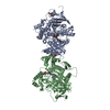
 Components Components | Acetylcholinesterase | ||||||
 Keywords Keywords | HYDROLASE / dual binding site inhibitor / chiral separation / acetylcholinesterase / Alzheimer's disease | ||||||
| Function / homology |  Function and homology information Function and homology informationacetylcholine catabolic process in synaptic cleft / acetylcholinesterase / choline metabolic process / acetylcholinesterase activity / side of membrane / synaptic cleft / synapse / : / plasma membrane Similarity search - Function | ||||||
| Biological species |   Tetronarce californica (Pacific electric ray) Tetronarce californica (Pacific electric ray) | ||||||
| Method |  X-RAY DIFFRACTION / SYNCHROTRON / MOLECULAR REPLACEMENT / Resolution: 2.80003021833 Å X-RAY DIFFRACTION / SYNCHROTRON / MOLECULAR REPLACEMENT / Resolution: 2.80003021833 Å | ||||||
 Authors Authors | de la Mora, E. / Mangiatordi, G.F. / Belviso, B.D. / Caliandro, R. / Colletier, J.P. / Catto, M. | ||||||
 Citation Citation | Journal: Acs Med.Chem.Lett. / Year: 2020 Title: Chiral Separation, X-ray Structure, and Biological Evaluation of a Potent and Reversible Dual Binding Site AChE Inhibitor. Authors: Catto, M. / Pisani, L. / de la Mora, E. / Belviso, B.D. / Mangiatordi, G.F. / Pinto, A. / Palma, A. / Denora, N. / Caliandro, R. / Colletier, J.P. / Silman, I. / Nicolotti, O. / Altomare, C.D. | ||||||
| History |
|
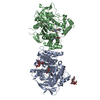
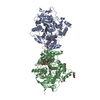
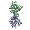
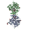
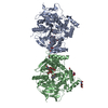
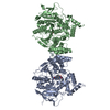
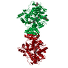
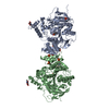
- Structure visualization
| Structure viewer | Molecule: MolmilJmol/JSmol |
|---|
- Downloads & links
-Download
| PDBx/mmCIF format | 6tt0.cif.gz | 135.5 KB | Display | PDBx/mmCIF format |
|---|---|---|---|---|
| PDB format | pdb6tt0.ent.gz | 94.1 KB | Display | PDB format |
| PDBx/mmJSON format | 6tt0.json.gz | Tree view | PDBx/mmJSON format | |
| Others |  Other downloads Other downloads |
-Validation report
| Arichive directory | https://data.pdbj.org/pub/pdb/validation_reports/tt/6tt0ftp://data.pdbj.org/pub/pdb/validation_reports/tt/6tt0 | HTTPS FTP |
|---|
-Related structure data
| Related structure data | 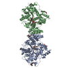 2vt7S S: Starting model for refinement |
|---|---|
| Similar structure data |


-Links
 PDBj
PDBj
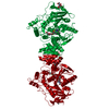
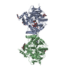
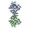
- Assembly
| Deposited unit | 
| ||||||||||||
|---|---|---|---|---|---|---|---|---|---|---|---|---|---|
| 1 | 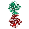
| ||||||||||||
| Unit cell |
|
-Components
| #1: Protein | Mass: 65315.594 Da / Num. of mol.: 1 Source method: isolated from a genetically manipulated source Details: T. californica acetylcholinesterase Source: (gene. exp.) Tetronarce californica (Pacific electric ray)Gene: ache Production host: Tetronarce californica (Pacific electric ray)References: UniProt: P04058, acetylcholinesterase | ||||||
|---|---|---|---|---|---|---|---|
| #2: Chemical | ChemComp-N9T / (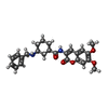  Mass: 436.500 Da / Num. of mol.: 1 / Source method: obtained synthetically / Formula: C25H28N2O5 / Feature type: SUBJECT OF INVESTIGATION Mass: 436.500 Da / Num. of mol.: 1 / Source method: obtained synthetically / Formula: C25H28N2O5 / Feature type: SUBJECT OF INVESTIGATION | ||||||
| #3: Sugar |   Type: D-saccharide, beta linking / Mass: 221.208 Da / Num. of mol.: 2 Type: D-saccharide, beta linking / Mass: 221.208 Da / Num. of mol.: 2Source method: isolated from a genetically manipulated source Formula: C8H15NO6 #4: Water | ChemComp-HOH / |  Mass: 18.015 Da / Num. of mol.: 88 / Source method: isolated from a natural source / Formula: H2O Mass: 18.015 Da / Num. of mol.: 88 / Source method: isolated from a natural source / Formula: H2OHas ligand of interest | Y | Has protein modification | Y | |
-Experimental details
-Experiment
| Experiment | Method: X-RAY DIFFRACTION / Number of used crystals: 1 |
|---|
- Sample preparation
| Crystal | Density Matthews: 4.1 Å3/Da / Density % sol: 70 % |
|---|---|
| Crystal grow | Temperature: 293 K / Method: vapor diffusion, hanging drop / pH: 5.5 / Details: 32% PEG 200 100 mM MES pH 5.5 |
-Data collection
| Diffraction | Mean temperature: 100 K / Serial crystal experiment: N |
|---|---|
| Diffraction source | Source: SYNCHROTRON / Site: ESRF  / Beamline: ID29 / Wavelength: 1.074 Å / Beamline: ID29 / Wavelength: 1.074 Å |
| Detector | Type: DECTRIS PILATUS 6M / Detector: PIXEL / Date: Aug 28, 2017 |
| Radiation | Protocol: SINGLE WAVELENGTH / Monochromatic (M) / Laue (L): M / Scattering type: x-ray |
| Radiation wavelength | Wavelength: 1.074 Å / Relative weight: 1 |
| Reflection | Resolution: 2.8→39.48 Å / Num. obs: 24409 / % possible obs: 98.1 % / Observed criterion σ(I): 1.5 / Redundancy: 1.8 % / Biso Wilson estimate: 67.6781327552 Å2 / CC1/2: 0.998 / Rmerge(I) obs: 0.04067 / Rpim(I) all: 0.04067 / Rrim(I) all: 0.05751 / Net I/σ(I): 10.5 |
| Reflection shell | Resolution: 2.8→2.9 Å / Redundancy: 1.8 % / Rmerge(I) obs: 0.3768 / Mean I/σ(I) obs: 1.57 / Num. unique obs: 4385 / CC1/2: 0.872 / Rsym value: 0.5328 / % possible all: 98.81 |
- Processing
| Software |
| |||||||||||||||||||||||||||||||||||||||||||||||||||||||||||||||
|---|---|---|---|---|---|---|---|---|---|---|---|---|---|---|---|---|---|---|---|---|---|---|---|---|---|---|---|---|---|---|---|---|---|---|---|---|---|---|---|---|---|---|---|---|---|---|---|---|---|---|---|---|---|---|---|---|---|---|---|---|---|---|---|---|
| Refinement | Method to determine structure: MOLECULAR REPLACEMENT Starting model: 2vt7 Resolution: 2.80003021833→39.4797998252 Å / SU ML: 0.395506018796 / Cross valid method: FREE R-VALUE / σ(F): 1.34889918222 / Phase error: 27.3771183945
| |||||||||||||||||||||||||||||||||||||||||||||||||||||||||||||||
| Solvent computation | Shrinkage radii: 0.9 Å / VDW probe radii: 1.11 Å | |||||||||||||||||||||||||||||||||||||||||||||||||||||||||||||||
| Displacement parameters | Biso mean: 69.9462107358 Å2 | |||||||||||||||||||||||||||||||||||||||||||||||||||||||||||||||
| Refinement step | Cycle: LAST / Resolution: 2.80003021833→39.4797998252 Å
| |||||||||||||||||||||||||||||||||||||||||||||||||||||||||||||||
| Refine LS restraints |
| |||||||||||||||||||||||||||||||||||||||||||||||||||||||||||||||
| LS refinement shell |
|
