 Movie
Movie Controller

[English] 日本語
 Yorodumi
- PDB-1ea5: NATIVE ACETYLCHOLINESTERASE (E.C. 3.1.1.7) FROM TORPEDO CALIFORNI... -
+ Open data

- Basic information
| Entry | Database: PDB / ID: 1ea5 | |||||||||
|---|---|---|---|---|---|---|---|---|---|---|
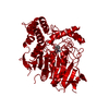
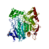
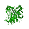
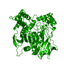
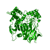
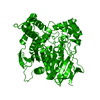
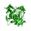
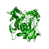
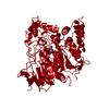
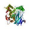
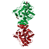
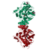
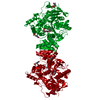
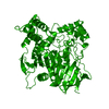
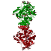
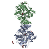
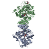
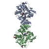
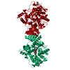
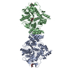
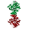
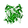
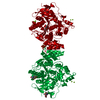
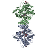
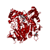
| Title | NATIVE ACETYLCHOLINESTERASE (E.C. 3.1.1.7) FROM TORPEDO CALIFORNICA at 1.8A resolution | |||||||||
 Components Components | ACETYLCHOLINESTERASE | |||||||||
 Keywords Keywords | HYDROLASE / SERINE HYDROLASE / NEUROTRANSMITTER CLEAVAGE / CATALYTIC TRIAD / ALPHA/BETA HYDROLASE | |||||||||
| Function / homology |  Function and homology information Function and homology informationacetylcholine catabolic process in synaptic cleft / acetylcholinesterase / choline metabolic process / acetylcholinesterase activity / side of membrane / synaptic cleft / synapse / : / plasma membrane Similarity search - Function | |||||||||
| Biological species |   TORPEDO CALIFORNICA (Pacific electric ray) TORPEDO CALIFORNICA (Pacific electric ray) | |||||||||
| Method |  X-RAY DIFFRACTION / SYNCHROTRON / MOLECULAR REPLACEMENT / Resolution: 1.8 Å X-RAY DIFFRACTION / SYNCHROTRON / MOLECULAR REPLACEMENT / Resolution: 1.8 Å | |||||||||
 Authors Authors | Harel, M. / Weik, M. / Silman, I. / Sussman, J.L. | |||||||||
 Citation Citation | Journal: Biochemistry / Year: 2002 Title: X-Ray Structures of Torpedo Californica Acetylcholinesterase Complexed with (+)-Huperzine a and (-)-Huperzine B: Structural Evidence for an Active Site Rearrangement Authors: Dvir, H. / Jiang, H.L. / Wong, D.M. / Harel, M. / Chetrit, M. / He, X.C. / Jin, G.Y. / Yu, G.L. / Tang, X.C. / Silman, I. / Bai, D.L. / Sussman, J.L. #1: Journal: Biochim.Biophys.Acta / Year: 1996 Title: Residues in Torpedo Californica Acetylcholinesterase Necessary for Processing to a Glycosyl Phosphatidylinositol-Anchored Form Authors: Bucht, G. / Hjalmarsson, K. #2: Journal: Protein Sci. / Year: 1994 Title: Structure and Dynamics of the Active Site Gorge of Acetylcholinesterase: Synergistic Use of Molecular Dynamics Simulation and X-Ray Crystallography Authors: Axelsen, P.H. / Harel, M. / Silman, I. / Sussman, J.L. #3: Journal: Proc.Natl.Acad.Sci.USA / Year: 1993Title: Quaternary Ligand Binding to Aromatic Residues in the Active-Site Gorge of Acetylcholinesterase Authors: Harel, M. / Schalk, I. / Ehret-Sabatier, L. / Bouet, F. / Goeldner, M. / Hirth, C. / Axelsen, P.H. / Silman, I. / Sussman, J.L. #4: Journal: Science / Year: 1991 Title: Atomic Structure of Acetylcholinesterase from Torpedo Californica: A Prototypic Acetylcholine-Binding Protein Authors: Sussman, J.L. / Harel, M. / Frolow, F. / Oefner, C. / Goldman, A. / Toker, L. / Silman, I. #5: Journal: J.Mol.Biol. / Year: 1988 Title: Purification and Crystallization of a Dimeric Form of Acetylcholinesterase from Torpedo Californica Subsequent to Solubilization with Phosphatidylinositol-Specific Phospholipase C Authors: Sussman, J.L. / Harel, M. / Frolow, F. / Varon, L. / Toker, L. / Futerman, A.H. / Silman, I. #6: Journal: Nature / Year: 1986 Title: Primary Structure of Torpedo Californica Acetylcholinesterase Deduced from its Cdna Sequence Authors: Schumacher, M. / Camp, S. / Maulet, Y. / Newton, M. / Macphee-Quigley, K. / Taylor, S.S. / Friedmann, T. / Taylor, P. | |||||||||
| History |
|
- Structure visualization
| Structure viewer | Molecule: MolmilJmol/JSmol |
|---|
- Downloads & links
-Download
| PDBx/mmCIF format | 1ea5.cif.gz | 141.3 KB | Display | PDBx/mmCIF format |
|---|---|---|---|---|
| PDB format | pdb1ea5.ent.gz | 110.5 KB | Display | PDB format |
| PDBx/mmJSON format | 1ea5.json.gz | Tree view | PDBx/mmJSON format | |
| Others |  Other downloads Other downloads |
-Validation report
| Arichive directory | https://data.pdbj.org/pub/pdb/validation_reports/ea/1ea5ftp://data.pdbj.org/pub/pdb/validation_reports/ea/1ea5 | HTTPS FTP |
|---|
-Related structure data
| Related structure data |  1gpkC  1gpnC  2aceS S: Starting model for refinement C: citing same article ( |
|---|---|
| Similar structure data |


-Links
 PDBj
PDBj
- Assembly
| Deposited unit | 
| ||||||||
|---|---|---|---|---|---|---|---|---|---|
| 1 | 
| ||||||||
| Unit cell |
| ||||||||
| Components on special symmetry positions |
| ||||||||
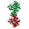
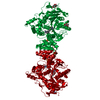
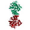
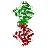
| Details | THE ENZYME IS A GPI-ANCHORED DIMER, THE TWO MONOMERS IN THE DIMER ARE RELATED BY CRYSTALLOGRAPHIC TWO-FOLD SYMMETRY. |
-Components
| #1: Protein | Mass: 60736.516 Da / Num. of mol.: 1 / Source method: isolated from a natural source Source: (natural) TORPEDO CALIFORNICA (Pacific electric ray)Organ: ELECTRIC ORGAN / Variant: G2 FORM / Tissue: ELECTROPLAQUE / References: UniProt: P04058, acetylcholinesterase | ||||||
|---|---|---|---|---|---|---|---|
| #2: Sugar |   Type: D-saccharide, beta linking / Mass: 221.208 Da / Num. of mol.: 2 Type: D-saccharide, beta linking / Mass: 221.208 Da / Num. of mol.: 2Source method: isolated from a genetically manipulated source Formula: C8H15NO6 #3: Water | ChemComp-HOH / |  Mass: 18.015 Da / Num. of mol.: 739 / Source method: isolated from a natural source / Formula: H2O Mass: 18.015 Da / Num. of mol.: 739 / Source method: isolated from a natural source / Formula: H2OCompound details | HYDROLYZES CHOLINE RELEASED INTO THE SYNAPSE. CATALYTIC ACTIVITY: ACETYLCHOLINE + H(2)O = CHOLINE + ...HYDROLYZES | Has protein modification | Y | |
-Experimental details
-Experiment
| Experiment | Method: X-RAY DIFFRACTION / Number of used crystals: 1 |
|---|
- Sample preparation
| Crystal | Density Matthews: 3.8 Å3/Da / Density % sol: 68 % |
|---|---|
| Crystal grow | Temperature: 277 K / pH: 5.8 Details: PROTEIN WAS CRYSTALLIZED FROM 35% PEG 200, 100 MM MES, PH 5.8, AT 4 DEG. |
-Data collection
| Diffraction | Mean temperature: 155 K |
|---|---|
| Diffraction source | Source: SYNCHROTRON / Site: ESRF  / Beamline: ID14-4 / Wavelength: 0.9312 / Beamline: ID14-4 / Wavelength: 0.9312 |
| Detector | Type: ADSC CCD / Detector: CCD / Date: Jun 15, 1999 |
| Radiation | Protocol: SINGLE WAVELENGTH / Monochromatic (M) / Laue (L): M / Scattering type: x-ray |
| Radiation wavelength | Wavelength: 0.9312 Å / Relative weight: 1 |
| Reflection | Resolution: 1.8→30 Å / Num. obs: 86444 / % possible obs: 92.9 % / Observed criterion σ(I): 0 / Redundancy: 1.9 % / Biso Wilson estimate: 33.9 Å2 / Rsym value: 0.067 / Net I/σ(I): 16.4 |
| Reflection shell | Resolution: 1.8→1.86 Å / Redundancy: 1.9 % / Mean I/σ(I) obs: 2.2 / Rsym value: 0.251 / % possible all: 68.8 |
- Processing
| Software |
| ||||||||||||||||||||||||||||||||||||||||||||||||||||||||||||||||||||||||||||||||
|---|---|---|---|---|---|---|---|---|---|---|---|---|---|---|---|---|---|---|---|---|---|---|---|---|---|---|---|---|---|---|---|---|---|---|---|---|---|---|---|---|---|---|---|---|---|---|---|---|---|---|---|---|---|---|---|---|---|---|---|---|---|---|---|---|---|---|---|---|---|---|---|---|---|---|---|---|---|---|---|---|---|
| Refinement | Method to determine structure: MOLECULAR REPLACEMENT Starting model: PDB ENTRY 2ACE Resolution: 1.8→27.34 Å / Rfactor Rfree error: 0.003 / Data cutoff high absF: 1738173.08 / Isotropic thermal model: RESTRAINED / Cross valid method: THROUGHOUT / σ(F): 0 Details: SEVERAL RESIDUES ARE NOT SEEN IN THE CRYSTAL STRUCTURE, DUE TO DISORDER. THESE INCLUDE THREE N- TERMINAL RESIDUES (ASP 1, ASP 2, HIS 3), AND THE C-TERMINAL RESIDUES AFTER THR 535. THERE ARE ...Details: SEVERAL RESIDUES ARE NOT SEEN IN THE CRYSTAL STRUCTURE, DUE TO DISORDER. THESE INCLUDE THREE N- TERMINAL RESIDUES (ASP 1, ASP 2, HIS 3), AND THE C-TERMINAL RESIDUES AFTER THR 535. THERE ARE 2 ALTERNATE CONFORMATIONS OBSERVED FOR RESIDUES: 9, 16, 19, 24, 46, 49, 55, 65, 221, 228, 260, 344, 353, 408, 430, 455, 468, 478, 508, 526.
| ||||||||||||||||||||||||||||||||||||||||||||||||||||||||||||||||||||||||||||||||
| Solvent computation | Solvent model: FLAT MODEL / Bsol: 82.8547 Å2 / ksol: 0.367212 e/Å3 | ||||||||||||||||||||||||||||||||||||||||||||||||||||||||||||||||||||||||||||||||
| Displacement parameters | Biso mean: 24.5 Å2
| ||||||||||||||||||||||||||||||||||||||||||||||||||||||||||||||||||||||||||||||||
| Refine analyze |
| ||||||||||||||||||||||||||||||||||||||||||||||||||||||||||||||||||||||||||||||||
| Refinement step | Cycle: LAST / Resolution: 1.8→27.34 Å
| ||||||||||||||||||||||||||||||||||||||||||||||||||||||||||||||||||||||||||||||||
| Refine LS restraints |
| ||||||||||||||||||||||||||||||||||||||||||||||||||||||||||||||||||||||||||||||||
| LS refinement shell | Resolution: 1.8→1.91 Å / Rfactor Rfree error: 0.012 / Total num. of bins used: 6
| ||||||||||||||||||||||||||||||||||||||||||||||||||||||||||||||||||||||||||||||||
| Xplor file |
|
