 Movie
Movie Controller


+ Open data

- Basic information
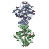
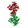
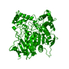
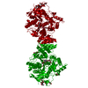
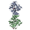
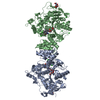
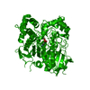
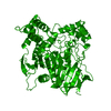
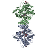
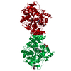
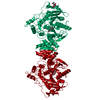
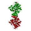
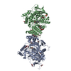
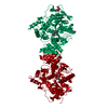
| Entry | Database: PDB / ID: 7b2w | ||||||
|---|---|---|---|---|---|---|---|
| Title | Torpedo californica acetylcholinesterase complexed with UO2 | ||||||
 Components Components | Acetylcholinesterase | ||||||
 Keywords Keywords | HYDROLASE / acetylcholinesterase / ASSAM / differential scanning calorimetry / divalent metal ion / electron paramagnetic resonance / thermal inactivation / Torpedo / 4D motif / 3.1.1.7 | ||||||
| Function / homology |  Function and homology information Function and homology informationacetylcholine catabolic process in synaptic cleft / acetylcholinesterase / choline metabolic process / acetylcholinesterase activity / side of membrane / synaptic cleft / synapse / : / plasma membrane Similarity search - Function | ||||||
| Biological species |   Tetronarce californica (Pacific electric ray) Tetronarce californica (Pacific electric ray) | ||||||
| Method |  X-RAY DIFFRACTION / MOLECULAR REPLACEMENT / Resolution: 2.65 Å X-RAY DIFFRACTION / MOLECULAR REPLACEMENT / Resolution: 2.65 Å | ||||||
 Authors Authors | Silman, I. / Shnyrov, V.L. / Ashani, Y. / Roth, E. / Nicolas, A. / Sussman, J.L. / Weiner, L. | ||||||
 Citation Citation | Journal: Protein Sci. / Year: 2021 Title: Torpedo californica acetylcholinesterase is stabilized by binding of a divalent metal ion to a novel and versatile 4D motif. Authors: Silman, I. / Shnyrov, V.L. / Ashani, Y. / Roth, E. / Nicolas, A. / Sussman, J.L. / Weiner, L. #1: Journal: Bull Soc Ophtalmol Fr / Year: 1987 Title: [Treatment of vitreous hemorrhages. Rare causes]. Authors: Aracil, P. #2: Journal: Science / Year: 1991 Title: Atomic structure of acetylcholinesterase from Torpedo californica: a prototypic acetylcholine-binding protein. Authors: Sussman, J.L. / Harel, M. / Frolow, F. / Oefner, C. / Goldman, A. / Toker, L. / Silman, I. | ||||||
| History |
|
- Structure visualization
| Structure viewer | Molecule: MolmilJmol/JSmol |
|---|
- Downloads & links
-Download
| PDBx/mmCIF format | 7b2w.cif.gz | 143.5 KB | Display | PDBx/mmCIF format |
|---|---|---|---|---|
| PDB format | pdb7b2w.ent.gz | 90.5 KB | Display | PDB format |
| PDBx/mmJSON format | 7b2w.json.gz | Tree view | PDBx/mmJSON format | |
| Others |  Other downloads Other downloads |
-Validation report
| Arichive directory | https://data.pdbj.org/pub/pdb/validation_reports/b2/7b2wftp://data.pdbj.org/pub/pdb/validation_reports/b2/7b2w | HTTPS FTP |
|---|
-Related structure data
| Related structure data | 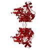 7b38C 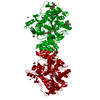 7b8eC 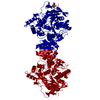 1ea5S S: Starting model for refinement C: citing same article ( |
|---|---|
| Similar structure data |


-Links
 PDBj
PDBj

- Assembly
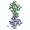
| Deposited unit | 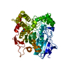
| ||||||||||||
|---|---|---|---|---|---|---|---|---|---|---|---|---|---|
| 1 | 
| ||||||||||||
| Unit cell |
|
-Components
| #1: Protein | Mass: 60736.516 Da / Num. of mol.: 1 / Source method: isolated from a natural source Source: (natural) Tetronarce californica (Pacific electric ray)Organ: ELECTRIC ORGAN / Variant: G2 FORM / Tissue: ELECTROPLAQUE / References: UniProt: P04058, acetylcholinesterase | ||||||
|---|---|---|---|---|---|---|---|
| #2: Chemical |   Mass: 270.028 Da / Num. of mol.: 2 / Source method: obtained synthetically / Formula: O2U / Feature type: SUBJECT OF INVESTIGATION Mass: 270.028 Da / Num. of mol.: 2 / Source method: obtained synthetically / Formula: O2U / Feature type: SUBJECT OF INVESTIGATION#3: Water | ChemComp-HOH / |  Mass: 18.015 Da / Num. of mol.: 27 / Source method: isolated from a natural source / Formula: H2O Mass: 18.015 Da / Num. of mol.: 27 / Source method: isolated from a natural source / Formula: H2OHas ligand of interest | Y | Has protein modification | Y | |
-Experimental details
-Experiment
| Experiment | Method: X-RAY DIFFRACTION / Number of used crystals: 1 |
|---|
- Sample preparation
| Crystal | Density Matthews: 3.93 Å3/Da / Density % sol: 68.74 % |
|---|---|
| Crystal grow | Temperature: 292 K / Method: vapor diffusion, hanging drop / pH: 7 Details: The use of standard vapor diffusion techniques in hanging drop with 61 percent saturated ammonium sulfate, 360 nM Na,K-phosphate buffer pH 7.0, as the precipating agent and a protein ...Details: The use of standard vapor diffusion techniques in hanging drop with 61 percent saturated ammonium sulfate, 360 nM Na,K-phosphate buffer pH 7.0, as the precipating agent and a protein concentration of ~11 mg/ml together with UO2(NO3)2. |
-Data collection
| Diffraction | Mean temperature: 293 K / Serial crystal experiment: N |
|---|---|
| Diffraction source | Source: ROTATING ANODE / Type: RIGAKU RU300 / Wavelength: 1.541 Å |
| Detector | Type: SIEMENS-XENTRONICS / Detector: AREA DETECTOR / Date: Jul 10, 1990 |
| Radiation | Monochromator: Graphite monochromator / Protocol: SINGLE WAVELENGTH / Monochromatic (M) / Laue (L): M / Scattering type: x-ray |
| Radiation wavelength | Wavelength: 1.541 Å / Relative weight: 1 |
| Reflection | Resolution: 2.65→39.0924 Å / Num. obs: 23867 / % possible obs: 84.24 % / Biso Wilson estimate: 36.6 Å2 / Rsym value: 0.115 / Net I/σ(I): 13.6 |
| Reflection shell | Resolution: 2.65→2.77 Å / Mean I/σ(I) obs: 1.44 / Num. unique obs: 2116 / Rsym value: 0.09 / % possible all: 57.9 |
- Processing
| Software |
| |||||||||||||||||||||||||||||||||||||||||||||||||||||||||||||||||||||||||||||||||||||||||||||||||||||||||
|---|---|---|---|---|---|---|---|---|---|---|---|---|---|---|---|---|---|---|---|---|---|---|---|---|---|---|---|---|---|---|---|---|---|---|---|---|---|---|---|---|---|---|---|---|---|---|---|---|---|---|---|---|---|---|---|---|---|---|---|---|---|---|---|---|---|---|---|---|---|---|---|---|---|---|---|---|---|---|---|---|---|---|---|---|---|---|---|---|---|---|---|---|---|---|---|---|---|---|---|---|---|---|---|---|---|---|
| Refinement | Method to determine structure: MOLECULAR REPLACEMENT Starting model: 1EA5 Resolution: 2.65→39.09 Å / SU ML: 0.3211 / Cross valid method: FREE R-VALUE / σ(F): 1.57 / Phase error: 20.046 Stereochemistry target values: GeoStd + Monomer Library + CDL v1.2
| |||||||||||||||||||||||||||||||||||||||||||||||||||||||||||||||||||||||||||||||||||||||||||||||||||||||||
| Solvent computation | Shrinkage radii: 0.9 Å / VDW probe radii: 1.11 Å / Solvent model: FLAT BULK SOLVENT MODEL | |||||||||||||||||||||||||||||||||||||||||||||||||||||||||||||||||||||||||||||||||||||||||||||||||||||||||
| Displacement parameters | Biso mean: 35.31 Å2 | |||||||||||||||||||||||||||||||||||||||||||||||||||||||||||||||||||||||||||||||||||||||||||||||||||||||||
| Refinement step | Cycle: LAST / Resolution: 2.65→39.09 Å
| |||||||||||||||||||||||||||||||||||||||||||||||||||||||||||||||||||||||||||||||||||||||||||||||||||||||||
| Refine LS restraints |
| |||||||||||||||||||||||||||||||||||||||||||||||||||||||||||||||||||||||||||||||||||||||||||||||||||||||||
| LS refinement shell |
|
