 Movie
Movie Controller


+ Open data

- Basic information
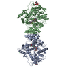
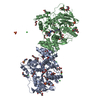
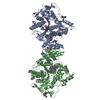
| Entry | Database: PDB / ID: 7b8e | ||||||
|---|---|---|---|---|---|---|---|
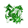
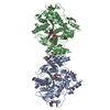
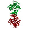
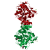
| Title | Torpedo californica acetylcholinesterase complexed with Ca+2 | ||||||
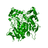
 Components Components | Acetylcholinesterase | ||||||
 Keywords Keywords | HYDROLASE / acetylcholinesterase / ASSAM / differential scanning calorimetry / divalent metal ion / electron paramagnetic resonance / thermal inactivation / Torpedo / 4D motif / 3.1.1.7 | ||||||
| Function / homology |  Function and homology information Function and homology informationacetylcholine catabolic process in synaptic cleft / acetylcholinesterase / choline metabolic process / acetylcholinesterase activity / side of membrane / synaptic cleft / synapse / : / plasma membrane Similarity search - Function | ||||||
| Biological species |   Tetronarce californica (Pacific electric ray) Tetronarce californica (Pacific electric ray) | ||||||
| Method |  X-RAY DIFFRACTION / SYNCHROTRON / MOLECULAR REPLACEMENT / Resolution: 2.23 Å X-RAY DIFFRACTION / SYNCHROTRON / MOLECULAR REPLACEMENT / Resolution: 2.23 Å | ||||||
 Authors Authors | Silman, I. / Shnyrov, V.L. / Ashani, Y. / Roth, E. / Nicolas, A. / Sussman, J.L. | ||||||
 Citation Citation | Journal: Protein Sci. / Year: 2021 Title: Torpedo californica acetylcholinesterase is stabilized by binding of a divalent metal ion to a novel and versatile 4D motif. Authors: Silman, I. / Shnyrov, V.L. / Ashani, Y. / Roth, E. / Nicolas, A. / Sussman, J.L. / Weiner, L. #1: Journal: Biochemistry / Year: 2002Title: X-ray structures of Torpedo californica acetylcholinesterase complexed with (+)-huperzine A and (-)-huperzine B: structural evidence for an active site rearrangement. Authors: Dvir, H. / Jiang, H.L. / Wong, D.M. / Harel, M. / Chetrit, M. / He, X.C. / Jin, G.Y. / Yu, G.L. / Tang, X.C. / Silman, I. / Bai, D.L. / Sussman, J.L. #2: Journal: Science / Year: 1991 Title: Atomic structure of acetylcholinesterase from Torpedo californica: a prototypic acetylcholine-binding protein. Authors: Sussman, J.L. / Harel, M. / Frolow, F. / Oefner, C. / Goldman, A. / Toker, L. / Silman, I. | ||||||
| History |
|
- Structure visualization
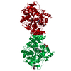
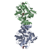
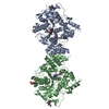
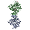
| Structure viewer | Molecule: MolmilJmol/JSmol |
|---|
- Downloads & links
-Download
| PDBx/mmCIF format | 7b8e.cif.gz | 157.1 KB | Display | PDBx/mmCIF format |
|---|---|---|---|---|
| PDB format | pdb7b8e.ent.gz | 98.1 KB | Display | PDB format |
| PDBx/mmJSON format | 7b8e.json.gz | Tree view | PDBx/mmJSON format | |
| Others |  Other downloads Other downloads |
-Validation report
| Arichive directory | https://data.pdbj.org/pub/pdb/validation_reports/b8/7b8eftp://data.pdbj.org/pub/pdb/validation_reports/b8/7b8e | HTTPS FTP |
|---|
-Related structure data
| Related structure data | 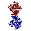 7b2wC  7b38C 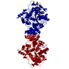 1ea5S S: Starting model for refinement C: citing same article ( |
|---|---|
| Similar structure data |


-Links
 PDBj
PDBj
- Assembly
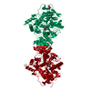
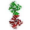
| Deposited unit | 
| ||||||||||||
|---|---|---|---|---|---|---|---|---|---|---|---|---|---|
| 1 | 
| ||||||||||||
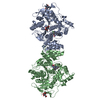
| Unit cell |
| ||||||||||||
| Components on special symmetry positions |
|
-Components
-Protein , 1 types, 1 molecules A
| #1: Protein | Mass: 60736.516 Da / Num. of mol.: 1 / Source method: isolated from a natural source Source: (natural) Tetronarce californica (Pacific electric ray)Organ: ELECTRIC ORGAN / Variant: G2 FORM / Tissue: ELECTROPLAQUE / References: UniProt: P04058, acetylcholinesterase |
|---|
-Sugars , 3 types, 3 molecules 
| #2: Polysaccharide | 2-acetamido-2-deoxy-beta-D-glucopyranose-(1-4)-2-acetamido-2-deoxy-beta-D-glucopyranose Source method: isolated from a genetically manipulated source |
|---|---|
| #3: Polysaccharide | alpha-L-fucopyranose-(1-6)-2-acetamido-2-deoxy-beta-D-glucopyranose Source method: isolated from a genetically manipulated source |
| #5: Sugar | ChemComp-NAG /  Type: D-saccharide, beta linking / Mass: 221.208 Da / Num. of mol.: 1 / Source method: obtained synthetically / Formula: C8H15NO6 Type: D-saccharide, beta linking / Mass: 221.208 Da / Num. of mol.: 1 / Source method: obtained synthetically / Formula: C8H15NO6 |
-Non-polymers , 5 types, 254 molecules 
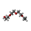


| #4: Chemical |  Mass: 62.068 Da / Num. of mol.: 3 / Source method: obtained synthetically / Formula: C2H6O2 Mass: 62.068 Da / Num. of mol.: 3 / Source method: obtained synthetically / Formula: C2H6O2#6: Chemical | ChemComp-FWN / |  Mass: 178.226 Da / Num. of mol.: 1 / Source method: obtained synthetically / Formula: C8H18O4 Mass: 178.226 Da / Num. of mol.: 1 / Source method: obtained synthetically / Formula: C8H18O4#7: Chemical | ChemComp-CA / |  Mass: 40.078 Da / Num. of mol.: 1 / Source method: obtained synthetically / Formula: Ca / Feature type: SUBJECT OF INVESTIGATION Mass: 40.078 Da / Num. of mol.: 1 / Source method: obtained synthetically / Formula: Ca / Feature type: SUBJECT OF INVESTIGATION#8: Chemical | ChemComp-ZN / |  Mass: 65.409 Da / Num. of mol.: 1 / Source method: obtained synthetically / Formula: Zn Mass: 65.409 Da / Num. of mol.: 1 / Source method: obtained synthetically / Formula: Zn#9: Water | ChemComp-HOH / | Mass: 18.015 Da / Num. of mol.: 248 / Source method: isolated from a natural source / Formula: H2O |
|---|
-Details
| Has ligand of interest | Y |
|---|---|
| Has protein modification | Y |
-Experimental details
-Experiment
| Experiment | Method: X-RAY DIFFRACTION / Number of used crystals: 1 |
|---|
- Sample preparation
| Crystal | Density Matthews: 3.3 Å3/Da / Density % sol: 62.7 % |
|---|---|
| Crystal grow | Temperature: 293 K / Method: vapor diffusion, hanging drop / pH: 5.8 Details: By mixing 2 uL of protein (10-13 mg/mL in 0.1 M NaCl/0.1 M 2-morpholinoethanesulfonic acid (MES)/0.02% sodium azide, pH 5.8) with 2 uL of precipitant solution (0.2 M calcium acetate/10-15% ...Details: By mixing 2 uL of protein (10-13 mg/mL in 0.1 M NaCl/0.1 M 2-morpholinoethanesulfonic acid (MES)/0.02% sodium azide, pH 5.8) with 2 uL of precipitant solution (0.2 M calcium acetate/10-15% (v/v) polyethylene glycol (PEG) 5000 monomethyl ether, 0.1 M MES, pH 6.5), thus yielding crystals of the Ca+2/TcAChE complex. |
-Data collection
| Diffraction | Mean temperature: 155 K / Serial crystal experiment: N |
|---|---|
| Diffraction source | Source: SYNCHROTRON / Site: ESRF  / Beamline: ID14-2 / Wavelength: 0.93 Å / Beamline: ID14-2 / Wavelength: 0.93 Å |
| Detector | Type: ADSC QUANTUM 1 / Detector: CCD / Date: Feb 10, 2000 |
| Radiation | Protocol: SINGLE WAVELENGTH / Monochromatic (M) / Laue (L): M / Scattering type: x-ray |
| Radiation wavelength | Wavelength: 0.93 Å / Relative weight: 1 |
| Reflection | Resolution: 2.23→19.96 Å / Num. obs: 38704 / % possible obs: 99.36 % / Redundancy: 4 % / Biso Wilson estimate: 39.68 Å2 / Rmerge(I) obs: 0.061 / Net I/σ(I): 11.6 |
| Reflection shell | Resolution: 2.25→2.33 Å / Rmerge(I) obs: 0.347 / Num. unique obs: 3752 |
- Processing
| Software |
| |||||||||||||||||||||||||||||||||||||||||||||||||||||||||||||||||||||||||||||||||||||||||||||||||||||||||
|---|---|---|---|---|---|---|---|---|---|---|---|---|---|---|---|---|---|---|---|---|---|---|---|---|---|---|---|---|---|---|---|---|---|---|---|---|---|---|---|---|---|---|---|---|---|---|---|---|---|---|---|---|---|---|---|---|---|---|---|---|---|---|---|---|---|---|---|---|---|---|---|---|---|---|---|---|---|---|---|---|---|---|---|---|---|---|---|---|---|---|---|---|---|---|---|---|---|---|---|---|---|---|---|---|---|---|
| Refinement | Method to determine structure: MOLECULAR REPLACEMENT Starting model: 1EA5 Resolution: 2.23→19.96 Å / SU ML: 0.2692 / Cross valid method: FREE R-VALUE / σ(F): 1.34 / Phase error: 24.1504 Stereochemistry target values: GeoStd + Monomer Library + CDL v1.2
| |||||||||||||||||||||||||||||||||||||||||||||||||||||||||||||||||||||||||||||||||||||||||||||||||||||||||
| Solvent computation | Shrinkage radii: 0.9 Å / VDW probe radii: 1.11 Å / Solvent model: FLAT BULK SOLVENT MODEL | |||||||||||||||||||||||||||||||||||||||||||||||||||||||||||||||||||||||||||||||||||||||||||||||||||||||||
| Displacement parameters | Biso mean: 43.68 Å2 | |||||||||||||||||||||||||||||||||||||||||||||||||||||||||||||||||||||||||||||||||||||||||||||||||||||||||
| Refinement step | Cycle: LAST / Resolution: 2.23→19.96 Å
| |||||||||||||||||||||||||||||||||||||||||||||||||||||||||||||||||||||||||||||||||||||||||||||||||||||||||
| Refine LS restraints |
| |||||||||||||||||||||||||||||||||||||||||||||||||||||||||||||||||||||||||||||||||||||||||||||||||||||||||
| LS refinement shell |
|
