 Movie
Movie Controller

[English] 日本語
 Yorodumi
- PDB-2bag: 3D Structure of Torpedo californica acetylcholinesterase complexe... -
+ Open data

- Basic information
| Entry | Database: PDB / ID: 2bag | ||||||
|---|---|---|---|---|---|---|---|
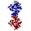
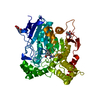
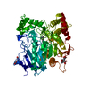
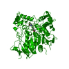
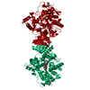
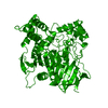
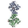
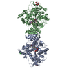
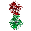
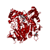
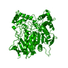
| Title | 3D Structure of Torpedo californica acetylcholinesterase complexed with Ganstigmine | ||||||
 Components Components | Acetylcholinesterase | ||||||
 Keywords Keywords | HYDROLASE / Serine hydrolase / Cholinesterase / Neurotransmitter cleavage / anti-Alzheimer drug | ||||||
| Function / homology |  Function and homology information Function and homology informationacetylcholine catabolic process in synaptic cleft / acetylcholinesterase / choline metabolic process / acetylcholinesterase activity / side of membrane / synaptic cleft / synapse / : / plasma membrane Similarity search - Function | ||||||
| Biological species |   Torpedo californica (Pacific electric ray) Torpedo californica (Pacific electric ray) | ||||||
| Method |  X-RAY DIFFRACTION / SYNCHROTRON / MOLECULAR REPLACEMENT / Resolution: 2.4 Å X-RAY DIFFRACTION / SYNCHROTRON / MOLECULAR REPLACEMENT / Resolution: 2.4 Å | ||||||
 Authors Authors | Lamba, D. / Bartolucci, C. / Siotto, M. / Racchi, M. / Villetti, G. / Delcanale, M. | ||||||
 Citation Citation | Journal: J.Med.Chem. / Year: 2006 Title: Structural Determinants of Torpedo californica Acetylcholinesterase Inhibition by the Novel and Orally Active Carbamate Based Anti-Alzheimer Drug Ganstigmine (CHF-2819) Authors: Bartolucci, C. / Siotto, M. / Ghidini, E. / Amari, G. / Bolzoni, P.T. / Racchi, M. / Villetti, G. / Delcanale, M. / Lamba, D. #1: Journal: Biochemistry / Year: 1999Title: Back Door opening implied by the crystal structure of a carbamoylated acetylcholinesterase Authors: Bartolucci, C. / Perola, E. / Cellai, L. / Brufani, M. / Lamba, D. #2: Journal: Biochemistry / Year: 2002Title: Kinetic and structural studies on the interaction of cholinesterases with the anti-Alzheimer drug rivastigmine Authors: Bar-On, P. / Millard, C.B. / Harel, M. / Dvir, H. / Enz, A. / Sussman, J.L. / Silman, I. #3: Journal: Biochim.Biophys.Acta / Year: 1997 Title: Long chain analogs of physostigmine as potential drugs for Alzheimer's disease: new insights into the mechanism of action in the inhibition of acetylcholinesterase Authors: Perola, E. / Cellai, L. / Lamba, D. / Filocamo, L. / Brufani, M. | ||||||
| History |
|
- Structure visualization
| Structure viewer | Molecule: MolmilJmol/JSmol |
|---|
- Downloads & links
-Download
| PDBx/mmCIF format | 2bag.cif.gz | 128.3 KB | Display | PDBx/mmCIF format |
|---|---|---|---|---|
| PDB format | pdb2bag.ent.gz | 97.6 KB | Display | PDB format |
| PDBx/mmJSON format | 2bag.json.gz | Tree view | PDBx/mmJSON format | |
| Others |  Other downloads Other downloads |
-Validation report
| Arichive directory | https://data.pdbj.org/pub/pdb/validation_reports/ba/2bagftp://data.pdbj.org/pub/pdb/validation_reports/ba/2bag | HTTPS FTP |
|---|
-Related structure data
| Related structure data | 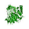 2aceS S: Starting model for refinement |
|---|---|
| Similar structure data |


-Links
 PDBj
PDBj
- Assembly
| Deposited unit | 
| ||||||||
|---|---|---|---|---|---|---|---|---|---|
| 1 | 
| ||||||||
| Unit cell |
|
-Components
-Protein / Sugars , 2 types, 3 molecules A
| #1: Protein | Mass: 61325.090 Da / Num. of mol.: 1 / Source method: isolated from a natural source Source: (natural) Torpedo californica (Pacific electric ray)Organ: Eletric organ / Variant: G2 form / Tissue: Electroplaque / References: UniProt: P04058, acetylcholinesterase |
|---|---|
| #2: Sugar |  Type: D-saccharide, beta linking / Mass: 221.208 Da / Num. of mol.: 2 Type: D-saccharide, beta linking / Mass: 221.208 Da / Num. of mol.: 2Source method: isolated from a genetically manipulated source Formula: C8H15NO6 |
-Non-polymers , 4 types, 278 molecules 


| #3: Chemical | ChemComp-1PE /  Mass: 238.278 Da / Num. of mol.: 1 / Source method: obtained synthetically / Formula: C10H22O6 / Comment: precipitant*YM Mass: 238.278 Da / Num. of mol.: 1 / Source method: obtained synthetically / Formula: C10H22O6 / Comment: precipitant*YM | ||
|---|---|---|---|
| #4: Chemical | ChemComp-GSG /  Mass: 381.468 Da / Num. of mol.: 1 / Source method: obtained synthetically / Formula: C22H27N3O3 Mass: 381.468 Da / Num. of mol.: 1 / Source method: obtained synthetically / Formula: C22H27N3O3 | ||
| #5: Chemical |  Mass: 195.237 Da / Num. of mol.: 2 / Source method: obtained synthetically / Formula: C6H13NO4S / Comment: pH buffer*YM Mass: 195.237 Da / Num. of mol.: 2 / Source method: obtained synthetically / Formula: C6H13NO4S / Comment: pH buffer*YM#6: Water | ChemComp-HOH / | Mass: 18.015 Da / Num. of mol.: 274 / Source method: isolated from a natural source / Formula: H2O |
-Details
| Has protein modification | Y |
|---|
-Experimental details
-Experiment
| Experiment | Method: X-RAY DIFFRACTION / Number of used crystals: 1 |
|---|
- Sample preparation
| Crystal | Density Matthews: 4 Å3/Da / Density % sol: 69.2 % |
|---|---|
| Crystal grow | Temperature: 277 K / Method: vapor diffusion, hanging drop / pH: 6 Details: 40% PEG200, 100mM MES, pH 6.0, VAPOR DIFFUSION, HANGING DROP, temperature 277K |
-Data collection
| Diffraction | Mean temperature: 100 K |
|---|---|
| Diffraction source | Source: SYNCHROTRON / Site: ELETTRA  / Beamline: 5.2R / Wavelength: 1 Å / Beamline: 5.2R / Wavelength: 1 Å |
| Detector | Type: MARRESEARCH / Detector: IMAGE PLATE / Date: May 31, 1998 / Details: Three-segment Pt-coated toroidal mirror |
| Radiation | Monochromator: Double Crystal (Si111) / Protocol: SINGLE WAVELENGTH / Monochromatic (M) / Laue (L): M / Scattering type: x-ray |
| Radiation wavelength | Wavelength: 1 Å / Relative weight: 1 |
| Reflection | Resolution: 2.4→27.93 Å / Num. all: 37817 / Num. obs: 37817 / % possible obs: 96.4 % / Observed criterion σ(F): -3 / Observed criterion σ(I): 0 / Redundancy: 6 % / Biso Wilson estimate: 35.3 Å2 / Rmerge(I) obs: 0.063 / Rsym value: 0.057 / Net I/σ(I): 8.6 |
| Reflection shell | Resolution: 2.4→2.44 Å / Redundancy: 5.1 % / Rmerge(I) obs: 0.47 / Mean I/σ(I) obs: 1.9 / Num. unique all: 2303 / Rsym value: 0.422 / % possible all: 97.4 |
- Processing
| Software |
| ||||||||||||||||||||||||||||||||||||||||||||||||||||||||||||
|---|---|---|---|---|---|---|---|---|---|---|---|---|---|---|---|---|---|---|---|---|---|---|---|---|---|---|---|---|---|---|---|---|---|---|---|---|---|---|---|---|---|---|---|---|---|---|---|---|---|---|---|---|---|---|---|---|---|---|---|---|---|
| Refinement | Method to determine structure: MOLECULAR REPLACEMENT Starting model: PDB ENTRY 2ACE Resolution: 2.4→27.93 Å / Rfactor Rfree error: 0.004 / Data cutoff high absF: 1599796.36 / Data cutoff low absF: 0 / Isotropic thermal model: Overall anisotropic / Cross valid method: THROUGHOUT / σ(F): 0 / σ(I): 0 / Stereochemistry target values: Engh & Huber
| ||||||||||||||||||||||||||||||||||||||||||||||||||||||||||||
| Solvent computation | Solvent model: Flat model / Bsol: 50.11 Å2 / ksol: 0.365 e/Å3 | ||||||||||||||||||||||||||||||||||||||||||||||||||||||||||||
| Displacement parameters | Biso mean: 45.6 Å2
| ||||||||||||||||||||||||||||||||||||||||||||||||||||||||||||
| Refine analyze |
| ||||||||||||||||||||||||||||||||||||||||||||||||||||||||||||
| Refinement step | Cycle: LAST / Resolution: 2.4→27.93 Å
| ||||||||||||||||||||||||||||||||||||||||||||||||||||||||||||
| Refine LS restraints |
| ||||||||||||||||||||||||||||||||||||||||||||||||||||||||||||
| LS refinement shell | Resolution: 2.4→2.55 Å / Rfactor Rfree error: 0.012 / Total num. of bins used: 6
|
