 Movie
Movie Controller

[English] 日本語
 Yorodumi
- PDB-3i6m: 3D Structure of Torpedo californica acetylcholinesterase complexe... -
+ Open data

- Basic information
| Entry | Database: PDB / ID: 3i6m | |||||||||
|---|---|---|---|---|---|---|---|---|---|---|
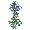
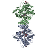
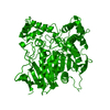
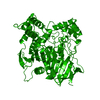
| Title | 3D Structure of Torpedo californica acetylcholinesterase complexed with N-piperidinopropyl-galanthamine | |||||||||
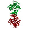
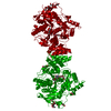
 Components Components | Acetylcholinesterase | |||||||||
 Keywords Keywords | HYDROLASE / Serine hydrolase / Cholinesterase / Neurotransmitter degradation / Alzheimer's disease / Bis-functional galanthamine derivative / Alternative splicing / Cell junction / Cell membrane / Disulfide bond / Glycoprotein / GPI-anchor / Lipoprotein / Membrane / Serine esterase / Synapse | |||||||||
| Function / homology |  Function and homology information Function and homology informationacetylcholine catabolic process in synaptic cleft / acetylcholinesterase / choline metabolic process / acetylcholinesterase activity / side of membrane / synaptic cleft / synapse / : / plasma membrane Similarity search - Function | |||||||||
| Biological species |   Torpedo californica (Pacific electric ray) Torpedo californica (Pacific electric ray) | |||||||||
| Method |  X-RAY DIFFRACTION / SYNCHROTRON / MOLECULAR REPLACEMENT / Resolution: 2.26 Å X-RAY DIFFRACTION / SYNCHROTRON / MOLECULAR REPLACEMENT / Resolution: 2.26 Å | |||||||||
 Authors Authors | Lamba, D. / Bartolucci, C. | |||||||||
 Citation Citation | Journal: J.Med.Chem. / Year: 2010 Title: Probing Torpedo californica acetylcholinesterase catalytic gorge with two novel bis-functional galanthamine derivatives. Authors: Bartolucci, C. / Haller, L.A. / Jordis, U. / Fels, G. / Lamba, D. #1: Journal: J.Am.Chem.Soc. / Year: 2004Title: The complex of a bivalent derivative of galanthamine with Torpedo acetylcholinesterase displays drastic deformation of the active-site gorge: implications for structure-based drug design Authors: Greenblatt, H.M. / Guillou, C. / Guenard, D. / Argaman, A. / Botti, S. / Badet, B. / Thal, C. / Silman, I. / Sussman, J.L. #2: Journal: FEBS Lett. / Year: 1999Title: Structure of acetylcholinesterase complexed with (-)-galanthamine at 2.3A resolution Authors: Greenblatt, H.M. / Kryger, G. / Lewis, T. / Silman, I. / Sussman, J.L. #3: Journal: PROTEINS / Year: 2001Title: Three-dimensional structure of a complex of galanthamine (Nivalin) with acetylcholinesterase from Torpedo californica: implications for the design of new anti-Alzheimer drugs Authors: Bartolucci, C. / Perola, E. / Pilger, C. / Fels, G. / Lamba, D. #4: Journal: J.MOL.GRAPH.MODEL. / Year: 2001 Title: Accurate prediction of the bound conformation of galanthamine in the active site of Torpedo californica acetylcholinesterase using molecular docking Authors: Pilger, C. / Bartolucci, C. / Lamba, D. / Tropsha, A. / Fels, G. #5: Journal: J.Mol.Model. / Year: 2002 Title: Galanthamine as bis-functional ligand for the acetylcholinesterase. Authors: Luttmann, E. / Linnemann, E. / Fels, G. #6: Journal: J.Chem.Inf.Model. / Year: 2006 Title: A QXP-based multistep docking procedure for accurate prediction of protein-ligand complexes Authors: Alisaraie, L. / Fels, G. | |||||||||
| History |
|
- Structure visualization
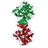
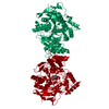
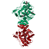
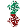
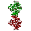
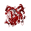
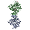
| Structure viewer | Molecule: MolmilJmol/JSmol |
|---|
- Downloads & links
-Download
| PDBx/mmCIF format | 3i6m.cif.gz | 132.5 KB | Display | PDBx/mmCIF format |
|---|---|---|---|---|
| PDB format | pdb3i6m.ent.gz | 100.5 KB | Display | PDB format |
| PDBx/mmJSON format | 3i6m.json.gz | Tree view | PDBx/mmJSON format | |
| Others |  Other downloads Other downloads |
-Validation report
| Arichive directory | https://data.pdbj.org/pub/pdb/validation_reports/i6/3i6mftp://data.pdbj.org/pub/pdb/validation_reports/i6/3i6m | HTTPS FTP |
|---|
-Related structure data
| Related structure data | 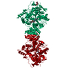 3i6zC 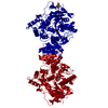 1ea5S C: citing same article ( S: Starting model for refinement |
|---|---|
| Similar structure data |


-Links
 PDBj
PDBj
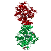
- Assembly
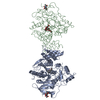
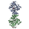
| Deposited unit | 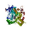
| ||||||||
|---|---|---|---|---|---|---|---|---|---|
| 1 | 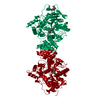
| ||||||||
| Unit cell |
|
-Components
| #1: Protein | Mass: 60447.211 Da / Num. of mol.: 1 / Fragment: UNP residues 23-556 / Source method: isolated from a natural source Source: (natural) Torpedo californica (Pacific electric ray)References: UniProt: P04058, acetylcholinesterase |
|---|---|
| #2: Polysaccharide | 2-acetamido-2-deoxy-beta-D-glucopyranose-(1-4)-2-acetamido-2-deoxy-beta-D-glucopyranose Source method: isolated from a genetically manipulated source |
| #3: Sugar | ChemComp-NAG /   Type: D-saccharide, beta linking / Mass: 221.208 Da / Num. of mol.: 1 Type: D-saccharide, beta linking / Mass: 221.208 Da / Num. of mol.: 1Source method: isolated from a genetically manipulated source Formula: C8H15NO6 |
| #4: Chemical | ChemComp-G3X / (  Mass: 398.538 Da / Num. of mol.: 1 / Source method: obtained synthetically / Formula: C24H34N2O3 Mass: 398.538 Da / Num. of mol.: 1 / Source method: obtained synthetically / Formula: C24H34N2O3 |
| #5: Water | ChemComp-HOH /  Mass: 18.015 Da / Num. of mol.: 398 / Source method: isolated from a natural source / Formula: H2O Mass: 18.015 Da / Num. of mol.: 398 / Source method: isolated from a natural source / Formula: H2O |
| Has protein modification | Y |
-Experimental details
-Experiment
| Experiment | Method: X-RAY DIFFRACTION / Number of used crystals: 1 |
|---|
- Sample preparation
| Crystal | Density Matthews: 4.12 Å3/Da / Density % sol: 70.17 % |
|---|---|
| Crystal grow | Temperature: 277 K / Method: vapor diffusion, hanging drop / pH: 6 Details: 42% PEG200, 100mM MES pH 6.0, VAPOR DIFFUSION, HANGING DROP, temperature 277K |
-Data collection
| Diffraction | Mean temperature: 120 K |
|---|---|
| Diffraction source | Source: SYNCHROTRON / Site: ELETTRA  / Beamline: 5.2R / Wavelength: 1 Å / Beamline: 5.2R / Wavelength: 1 Å |
| Detector | Type: MAR CCD 165 mm / Detector: CCD / Date: Apr 1, 2001 / Details: Three-segment Pt-coated toroidal mirrors |
| Radiation | Monochromator: Double crystal Si(111) / Protocol: SINGLE WAVELENGTH / Monochromatic (M) / Laue (L): M / Scattering type: x-ray |
| Radiation wavelength | Wavelength: 1 Å / Relative weight: 1 |
| Reflection | Resolution: 2.26→19.82 Å / Num. all: 47210 / Num. obs: 46878 / % possible obs: 99.7 % / Observed criterion σ(F): 0 / Observed criterion σ(I): -3 / Redundancy: 9.3 % / Biso Wilson estimate: 30.5 Å2 / Rsym value: 0.047 / Net I/σ(I): 8.8 |
| Reflection shell | Resolution: 2.26→2.28 Å / Redundancy: 2.9 % / Mean I/σ(I) obs: 2 / Num. unique all: 957 / Rsym value: 0.418 / % possible all: 97.3 |
- Processing
| Software |
| ||||||||||||||||||||||||||||||||||||
|---|---|---|---|---|---|---|---|---|---|---|---|---|---|---|---|---|---|---|---|---|---|---|---|---|---|---|---|---|---|---|---|---|---|---|---|---|---|
| Refinement | Method to determine structure: MOLECULAR REPLACEMENT Starting model: PDB ENTRY 1EA5 Resolution: 2.26→19.82 Å / Rfactor Rfree error: 0.003 / Data cutoff high absF: 1997132.14 / Isotropic thermal model: Restrained individual isotropic / Cross valid method: THROUGHOUT / σ(F): 0 / Stereochemistry target values: Engh & Huber
| ||||||||||||||||||||||||||||||||||||
| Solvent computation | Solvent model: FLAT MODEL / Bsol: 62.8 Å2 / ksol: 0.4 e/Å3 | ||||||||||||||||||||||||||||||||||||
| Displacement parameters | Biso mean: 40.2 Å2
| ||||||||||||||||||||||||||||||||||||
| Refine analyze |
| ||||||||||||||||||||||||||||||||||||
| Refinement step | Cycle: LAST / Resolution: 2.26→19.82 Å
| ||||||||||||||||||||||||||||||||||||
| Refine LS restraints |
| ||||||||||||||||||||||||||||||||||||
| LS refinement shell | Resolution: 2.26→2.33 Å / Rfactor Rfree error: 0.014 / Total num. of bins used: 6
|
