 Movie
Movie Controller

[English] 日本語
 Yorodumi
- PDB-1qif: SPECIFIC CHEMICAL AND STRUCTURAL DAMAGE AT NINE TIME POINTS (POIN... -
+ Open data

- Basic information
| Entry | Database: PDB / ID: 1qif | ||||||
|---|---|---|---|---|---|---|---|
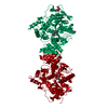
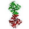
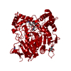
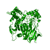
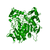
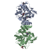
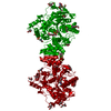
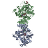
| Title | SPECIFIC CHEMICAL AND STRUCTURAL DAMAGE AT NINE TIME POINTS (POINT C) CAUSED BY INTENSE SYNCHROTRON RADIATION TO TORPEDO CALIFORNICA ACETYLCHOLINESTERASE | ||||||
 Components Components | ACETYLCHOLINESTERASE | ||||||
 Keywords Keywords | HYDROLASE / RADIATION DAMAGE / TIME SERIES / DISULFIDE BOND / SERINE HYDROLASE / ALPHA/BETA HYDROLASE / NEUROTRANSMITTER CLEAVAGE / CATALYTIC TRIAD / GLYCOSYLATED PROTEIN | ||||||
| Function / homology |  Function and homology information Function and homology informationacetylcholine catabolic process in synaptic cleft / acetylcholinesterase / choline metabolic process / acetylcholinesterase activity / side of membrane / synaptic cleft / synapse / : / plasma membrane Similarity search - Function | ||||||
| Biological species |   Torpedo californica (Pacific electric ray) Torpedo californica (Pacific electric ray) | ||||||
| Method |  X-RAY DIFFRACTION / SYNCHROTRON / MOLECULAR REPLACEMENT / Resolution: 2.1 Å X-RAY DIFFRACTION / SYNCHROTRON / MOLECULAR REPLACEMENT / Resolution: 2.1 Å | ||||||
 Authors Authors | Kryger, G. / Weik, M. / Ravelli, R.B.G. | ||||||
 Citation Citation | Journal: Proc.Natl.Acad.Sci.USA / Year: 2000 Title: Specific chemical and structural damage to proteins produced by synchrotron radiation. Authors: Weik, M. / Ravelli, R.B. / Kryger, G. / McSweeney, S. / Raves, M.L. / Harel, M. / Gros, P. / Silman, I. / Kroon, J. / Sussman, J.L. | ||||||
| History |
|
- Structure visualization
| Structure viewer | Molecule: MolmilJmol/JSmol |
|---|
- Downloads & links
-Download
| PDBx/mmCIF format | 1qif.cif.gz | 114.9 KB | Display | PDBx/mmCIF format |
|---|---|---|---|---|
| PDB format | pdb1qif.ent.gz | 89.2 KB | Display | PDB format |
| PDBx/mmJSON format | 1qif.json.gz | Tree view | PDBx/mmJSON format | |
| Others |  Other downloads Other downloads |
-Validation report
| Arichive directory | https://data.pdbj.org/pub/pdb/validation_reports/qi/1qifftp://data.pdbj.org/pub/pdb/validation_reports/qi/1qif | HTTPS FTP |
|---|
-Related structure data
| Related structure data |  1qidC  1qieC  1qigC  1qihC  1qiiC  1qijC  1qikC  1qimC 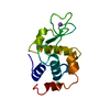 1qioC 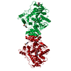 1vxrS C: citing same article ( S: Starting model for refinement |
|---|---|
| Similar structure data |


-Links
 PDBj
PDBj
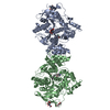
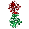
- Assembly
| Deposited unit | 
| ||||||||
|---|---|---|---|---|---|---|---|---|---|
| 1 | 
| ||||||||
| Unit cell |
|
-Components
| #1: Protein | Mass: 60736.516 Da / Num. of mol.: 1 / Source method: isolated from a natural source Source: (natural) Torpedo californica (Pacific electric ray)Organ: ELECTRIC ORGAN / Variant: G2 FORM / Tissue: ELECTROPLAQUE / References: UniProt: P04058, acetylcholinesterase |
|---|---|
| #2: Water | ChemComp-HOH /  Mass: 18.015 Da / Num. of mol.: 314 / Source method: isolated from a natural source / Formula: H2O Mass: 18.015 Da / Num. of mol.: 314 / Source method: isolated from a natural source / Formula: H2O |
-Experimental details
-Experiment
| Experiment | Method: X-RAY DIFFRACTION / Number of used crystals: 1 |
|---|
- Sample preparation
| Crystal | Density Matthews: 3.8 Å3/Da / Density % sol: 68 % | ||||||||||||||||||||
|---|---|---|---|---|---|---|---|---|---|---|---|---|---|---|---|---|---|---|---|---|---|
| Crystal grow | Temperature: 292 K / pH: 5.8 / Details: 30% PEG 200, 0.3 M MES, pH 5.8, temperature 292K | ||||||||||||||||||||
| Crystal grow | *PLUS Temperature: 4 ℃ / Method: vapor diffusion, hanging drop | ||||||||||||||||||||
| Components of the solutions | *PLUS
|
-Data collection
| Diffraction | Mean temperature: 100 K |
|---|---|
| Diffraction source | Source: SYNCHROTRON / Site: ESRF  / Beamline: ID14-4 / Wavelength: 0.932 / Beamline: ID14-4 / Wavelength: 0.932 |
| Detector | Type: ADSC QUANTUM 4 / Detector: CCD / Date: Mar 1, 1999 |
| Radiation | Protocol: SINGLE WAVELENGTH / Monochromatic (M) / Laue (L): M / Scattering type: x-ray |
| Radiation wavelength | Wavelength: 0.932 Å / Relative weight: 1 |
| Reflection | Resolution: 2.1→35.5 Å / Num. obs: 58334 / % possible obs: 99 % / Observed criterion σ(I): 0 / Redundancy: 1.96 % / Biso Wilson estimate: 23.5 Å2 / Rsym value: 4.5 |
| Reflection | *PLUS Lowest resolution: 40 Å / Redundancy: 1.8 % / Rmerge(I) obs: 0.052 |
| Reflection shell | *PLUS % possible obs: 99.2 % / Mean I/σ(I) obs: 1.6 |
- Processing
| Software |
| ||||||||||||||||||||||||||||||||||||||||||||||||||||||||||||||||||||||||||||||||
|---|---|---|---|---|---|---|---|---|---|---|---|---|---|---|---|---|---|---|---|---|---|---|---|---|---|---|---|---|---|---|---|---|---|---|---|---|---|---|---|---|---|---|---|---|---|---|---|---|---|---|---|---|---|---|---|---|---|---|---|---|---|---|---|---|---|---|---|---|---|---|---|---|---|---|---|---|---|---|---|---|---|
| Refinement | Method to determine structure: MOLECULAR REPLACEMENT Starting model: PDB ENTRY 1VXR Resolution: 2.1→35.5 Å / Rfactor Rfree error: 0.004 / Data cutoff high rms absF: 1834108.93 / Isotropic thermal model: RESTRAINED / Cross valid method: THROUGHOUT / σ(F): 0 Details: BULK SOLVENT MODEL USED. ONLY PARTIAL REFINEMENT DONE. ALL SIX CYSTEINE RESIDUES TAKING PART IN INTRACHAIN DISULFIDE LINKAGES,CYS 67-CYS 94, CYS 402- CYS 521 AND CYS 254-CYS 265, WERE ...Details: BULK SOLVENT MODEL USED. ONLY PARTIAL REFINEMENT DONE. ALL SIX CYSTEINE RESIDUES TAKING PART IN INTRACHAIN DISULFIDE LINKAGES,CYS 67-CYS 94, CYS 402- CYS 521 AND CYS 254-CYS 265, WERE MODELED AND REFINED AS ALANINE RESIDUES. TCACHE IS A GLYCOPROTEIN OF MR = 65,000, WHICH CONTAINS THREE INTRACHAIN DISULFIDE BONDS. IN THE COURSE OF CRYOGENIC DATA COLLECTION ON TRIGONAL CRYSTALS OF TCACHE ON THE UNDULATOR BEAMLINE, ID14-EH4, AT THE ESRF IN GRENOBLE, IN PREPARATION FOR TIME-RESOLVED STUDIES, WE COLLECTED A SERIES OF NINE HIGH-QUALITY COMPLETE DATA SETS ON THE SAME CRYSTAL. DATA COLLECTION UTILIZED THE UNATTENUATED BEAM, AND THE DURATION PER DATA SET WAS CA. 19 MIN, FOR 3 H IN TOTAL, AT 100K. ELECTRON DENSITY MAPS WERE OBTAINED FOR EACH DATA SET BY ROUTINE REFINEMENT, STARTING FROM THE SAME MODEL OF NATIVE TCACHE. FOR RESULTS, SEE: HTTP://SGJS3.WEIZMANN.AC.IL/~KRYGER/RADIATION_DAMAGE (LOWER CASE!) THIS ENTRY IS THE THIRD TIME POINT (3 OF 9). SEE HTTP:// SGJS3.WEIZMANN.AC.IL/~KRYGER/RADIATION_DAMAGE (LOWER CASE!)
| ||||||||||||||||||||||||||||||||||||||||||||||||||||||||||||||||||||||||||||||||
| Solvent computation | Solvent model: FLAT MODEL / Bsol: 56.7 Å2 / ksol: 0.365 e/Å3 | ||||||||||||||||||||||||||||||||||||||||||||||||||||||||||||||||||||||||||||||||
| Displacement parameters | Biso mean: 39.1 Å2
| ||||||||||||||||||||||||||||||||||||||||||||||||||||||||||||||||||||||||||||||||
| Refine analyze |
| ||||||||||||||||||||||||||||||||||||||||||||||||||||||||||||||||||||||||||||||||
| Refinement step | Cycle: LAST / Resolution: 2.1→35.5 Å
| ||||||||||||||||||||||||||||||||||||||||||||||||||||||||||||||||||||||||||||||||
| Refine LS restraints |
| ||||||||||||||||||||||||||||||||||||||||||||||||||||||||||||||||||||||||||||||||
| LS refinement shell | Resolution: 2.1→2.18 Å / Rfactor Rfree error: 0.018 / Total num. of bins used: 10
| ||||||||||||||||||||||||||||||||||||||||||||||||||||||||||||||||||||||||||||||||
| Xplor file |
| ||||||||||||||||||||||||||||||||||||||||||||||||||||||||||||||||||||||||||||||||
| Software | *PLUS Name: CNS / Version: 0.5 / Classification: refinement | ||||||||||||||||||||||||||||||||||||||||||||||||||||||||||||||||||||||||||||||||
| Refine LS restraints | *PLUS
|
