 Movie
Movie Controller


+ Open data

- Basic information
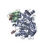
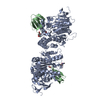
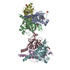
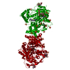
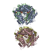
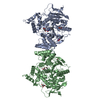
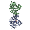
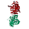
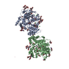
| Entry | Database: PDB / ID: 1fss | ||||||
|---|---|---|---|---|---|---|---|
| Title | ACETYLCHOLINESTERASE (E.C. 3.1.1.7) COMPLEXED WITH FASCICULIN-II | ||||||
 Components Components |
| ||||||
 Keywords Keywords | COMPLEX (SERINE ESTERASE/TOXIN) / COMPLEX (SERINE ESTERASE-TOXIN) / COMPLEX (SERINE ESTERASE-TOXIN) complex | ||||||
| Function / homology |  Function and homology information Function and homology informationacetylcholine catabolic process in synaptic cleft / acetylcholinesterase / choline metabolic process / acetylcholinesterase activity / side of membrane / synaptic cleft / toxin activity / synapse / : / extracellular region / plasma membrane Similarity search - Function | ||||||
| Biological species |   Torpedo californica (Pacific electric ray) Torpedo californica (Pacific electric ray) Dendroaspis angusticeps (eastern green mamba) Dendroaspis angusticeps (eastern green mamba) | ||||||
| Method |  X-RAY DIFFRACTION / SYNCHROTRON / Resolution: 3 Å X-RAY DIFFRACTION / SYNCHROTRON / Resolution: 3 Å | ||||||
 Authors Authors | Harel, M. / Kleywegt, G.J. / Silman, I. / Sussman, J.L. | ||||||
 Citation Citation | Journal: Structure / Year: 1995 Title: Crystal structure of an acetylcholinesterase-fasciculin complex: interaction of a three-fingered toxin from snake venom with its target. Authors: Harel, M. / Kleywegt, G.J. / Ravelli, R.B. / Silman, I. / Sussman, J.L. #1: Journal: J.Biol.Chem. / Year: 1992Title: 1.9-Angstroms Resolution Structure of Fasciculin 1, an Anti-Acetylcholine Esterase Toxin from Green Mamba Snake Venom Authors: Le Du, M.H. / Marchot, P. / Bougis, P.E. / Fontecilla-Camps, J.C. #2: Journal: Science / Year: 1991Title: Atomic Structure of Acetylcholine Esterase from Torpedo Californica: A Prototypic Acetylcholine Binding Enzyme Authors: Sussman, J.L. / Harel, M. / Frolow, F. / Oefner, C. / Goldman, A. / Toker, L. / Silman, I. | ||||||
| History |
|
- Structure visualization
| Structure viewer | Molecule: MolmilJmol/JSmol |
|---|
- Downloads & links
-Download
| PDBx/mmCIF format | 1fss.cif.gz | 117.2 KB | Display | PDBx/mmCIF format |
|---|---|---|---|---|
| PDB format | pdb1fss.ent.gz | 90.1 KB | Display | PDB format |
| PDBx/mmJSON format | 1fss.json.gz | Tree view | PDBx/mmJSON format | |
| Others |  Other downloads Other downloads |
-Validation report
| Arichive directory | https://data.pdbj.org/pub/pdb/validation_reports/fs/1fssftp://data.pdbj.org/pub/pdb/validation_reports/fs/1fss | HTTPS FTP |
|---|
-Related structure data
| Similar structure data |
|---|

-Links
 PDBj
PDBj

- Assembly
| Deposited unit | 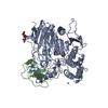
| ||||||||
|---|---|---|---|---|---|---|---|---|---|
| 1 | 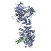
| ||||||||
| Unit cell |
| ||||||||
| Atom site foot note | 1: CIS PROLINE - PRO A 104 / 2: CIS PROLINE - PRO B 31 / 3: CIS PROLINE - PRO B 56 | ||||||||
| Components on special symmetry positions |
|
-Components
| #1: Protein | Mass: 60736.516 Da / Num. of mol.: 1 / Source method: isolated from a natural source Source: (natural) Torpedo californica (Pacific electric ray)Organ: ELECTRIC ORGAN / Variant: G2 FORM / Tissue: ELECTROPLAQUE / References: UniProt: P04058, acetylcholinesterase | ||||
|---|---|---|---|---|---|
| #2: Protein | Mass: 6768.769 Da / Num. of mol.: 1 / Source method: isolated from a natural source Source: (natural) Dendroaspis angusticeps (eastern green mamba)Organ: ELECTRIC ORGAN / Tissue: VENOM / References: UniProt: P01403, UniProt: P0C1Z0*PLUS | ||||
| #3: Sugar | ChemComp-NAG /   Type: D-saccharide, beta linking / Mass: 221.208 Da / Num. of mol.: 1 Type: D-saccharide, beta linking / Mass: 221.208 Da / Num. of mol.: 1Source method: isolated from a genetically manipulated source Formula: C8H15NO6 | ||||
| #4: Chemical |   Mass: 65.409 Da / Num. of mol.: 2 / Source method: obtained synthetically / Formula: Zn Mass: 65.409 Da / Num. of mol.: 2 / Source method: obtained synthetically / Formula: Zn#5: Water | ChemComp-HOH / |  Mass: 18.015 Da / Num. of mol.: 36 / Source method: isolated from a natural source / Formula: H2O Mass: 18.015 Da / Num. of mol.: 36 / Source method: isolated from a natural source / Formula: H2OHas protein modification | Y | |
-Experimental details
-Experiment
| Experiment | Method: X-RAY DIFFRACTION |
|---|
- Sample preparation
| Crystal | Density Matthews: 2.51 Å3/Da / Density % sol: 50.99 % | ||||||||||||||||||||||||||||||
|---|---|---|---|---|---|---|---|---|---|---|---|---|---|---|---|---|---|---|---|---|---|---|---|---|---|---|---|---|---|---|---|
| Crystal grow | *PLUS Temperature: 4 ℃ / pH: 5.2 / Method: vapor diffusionDetails: drop solution was mixed with an equal volume of reservoir solution | ||||||||||||||||||||||||||||||
| Components of the solutions | *PLUS
|
-Data collection
| Diffraction source | Source: SYNCHROTRON / Site: NSLS  / Beamline: X12C / Wavelength: 1.15 / Beamline: X12C / Wavelength: 1.15 |
|---|---|
| Detector | Type: MAR scanner 300 mm plate / Detector: IMAGE PLATE / Date: Nov 15, 1994 |
| Radiation | Monochromatic (M) / Laue (L): M / Scattering type: x-ray |
| Radiation wavelength | Wavelength: 1.15 Å / Relative weight: 1 |
| Reflection | Num. obs: 14206 / % possible obs: 97.7 % / Redundancy: 3.43 % / Rmerge(I) obs: 0.105 |
| Reflection | *PLUS Highest resolution: 3 Å / Num. measured all: 48744 / Rmerge(I) obs: 0.105 |
| Reflection shell | *PLUS Highest resolution: 3 Å / Lowest resolution: 3.11 Å / % possible obs: 93.7 % / Rmerge(I) obs: 0.303 / Mean I/σ(I) obs: 3.5 |
- Processing
| Software |
| ||||||||||||||||||||||||||||||||||||||||||||||||||||||||||||
|---|---|---|---|---|---|---|---|---|---|---|---|---|---|---|---|---|---|---|---|---|---|---|---|---|---|---|---|---|---|---|---|---|---|---|---|---|---|---|---|---|---|---|---|---|---|---|---|---|---|---|---|---|---|---|---|---|---|---|---|---|---|
| Refinement | Resolution: 3→8 Å / σ(F): 0
| ||||||||||||||||||||||||||||||||||||||||||||||||||||||||||||
| Displacement parameters | Biso mean: 19.6 Å2 | ||||||||||||||||||||||||||||||||||||||||||||||||||||||||||||
| Refinement step | Cycle: LAST / Resolution: 3→8 Å
| ||||||||||||||||||||||||||||||||||||||||||||||||||||||||||||
| Refine LS restraints |
| ||||||||||||||||||||||||||||||||||||||||||||||||||||||||||||
| Software | *PLUS Name: X-PLOR / Classification: refinement | ||||||||||||||||||||||||||||||||||||||||||||||||||||||||||||
| Refinement | *PLUS | ||||||||||||||||||||||||||||||||||||||||||||||||||||||||||||
| Solvent computation | *PLUS | ||||||||||||||||||||||||||||||||||||||||||||||||||||||||||||
| Displacement parameters | *PLUS | ||||||||||||||||||||||||||||||||||||||||||||||||||||||||||||
| Refine LS restraints | *PLUS
|
