 Movie
Movie Controller


+ Open data

- Basic information
| Entry | Database: PDB / ID: 1ax9 | ||||||
|---|---|---|---|---|---|---|---|
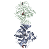
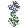
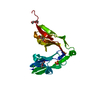
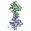
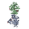
| Title | ACETYLCHOLINESTERASE COMPLEXED WITH EDROPHONIUM, LAUE DATA | ||||||
 Components Components | ACETYLCHOLINESTERASE | ||||||
 Keywords Keywords | HYDROLASE / CARBOXYLIC ESTERASE / SERINE ESTERASE / SYNAPSE | ||||||
| Function / homology |  Function and homology information Function and homology informationacetylcholine catabolic process in synaptic cleft / acetylcholinesterase / choline metabolic process / acetylcholinesterase activity / side of membrane / synaptic cleft / synapse / : / plasma membrane Similarity search - Function | ||||||
| Biological species |   Torpedo californica (Pacific electric ray) Torpedo californica (Pacific electric ray) | ||||||
| Method |  X-RAY DIFFRACTION / SYNCHROTRON / MOLECULAR REPLACEMENT / Resolution: 2.8 Å X-RAY DIFFRACTION / SYNCHROTRON / MOLECULAR REPLACEMENT / Resolution: 2.8 Å | ||||||
 Authors Authors | Raves, M.L. / Ravelli, R.B.G. / Sussman, J.L. / Harel, M. / Silman, I. | ||||||
 Citation Citation | Journal: Acta Crystallogr.,Sect.D / Year: 1998 Title: Static Laue diffraction studies on acetylcholinesterase. Authors: Ravelli, R.B. / Raves, M.L. / Ren, Z. / Bourgeois, D. / Roth, M. / Kroon, J. / Silman, I. / Sussman, J.L. #1: Journal: Proc.Natl.Acad.Sci.USA / Year: 1993Title: Quaternary Ligand Binding to Aromatic Residues in the Active-Site Gorge of Acetylcholinesterase Authors: Harel, M. / Schalk, I. / Ehret-Sabatier, L. / Bouet, F. / Goeldner, M. / Hirth, C. / Axelsen, P.H. / Silman, I. / Sussman, J.L. #2: Journal: Science / Year: 1991Title: Atomic Structure of Acetylcholinesterase from Torpedo Californica: A Prototypic Acetylcholine-Binding Protein Authors: Sussman, J.L. / Harel, M. / Frolow, F. / Oefner, C. / Goldman, A. / Toker, L. / Silman, I. #3: Journal: J.Mol.Biol. / Year: 1988Title: Purification and Crystallization of a Dimeric Form of Acetylcholinesterase from Torpedo Californica Subsequent to Solubilization with Phosphatidylinositol-Specific Phospholipase C Authors: Sussman, J.L. / Harel, M. / Frolow, F. / Varon, L. / Toker, L. / Futerman, A.H. / Silman, I. | ||||||
| History |
|
- Structure visualization
| Structure viewer | Molecule: MolmilJmol/JSmol |
|---|
- Downloads & links
-Download
| PDBx/mmCIF format | 1ax9.cif.gz | 117.1 KB | Display | PDBx/mmCIF format |
|---|---|---|---|---|
| PDB format | pdb1ax9.ent.gz | 90.3 KB | Display | PDB format |
| PDBx/mmJSON format | 1ax9.json.gz | Tree view | PDBx/mmJSON format | |
| Others |  Other downloads Other downloads |
-Validation report
| Arichive directory | https://data.pdbj.org/pub/pdb/validation_reports/ax/1ax9ftp://data.pdbj.org/pub/pdb/validation_reports/ax/1ax9 | HTTPS FTP |
|---|
-Related structure data
| Related structure data |  2ackC  2aceS S: Starting model for refinement C: citing same article ( |
|---|---|
| Similar structure data |


-Links
 PDBj
PDBj
- Assembly
| Deposited unit | 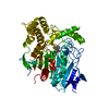
| ||||||||
|---|---|---|---|---|---|---|---|---|---|
| 1 | 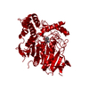
| ||||||||
| Unit cell |
|
-Components
| #1: Protein | Mass: 60736.516 Da / Num. of mol.: 1 / Source method: isolated from a natural source Source: (natural) Torpedo californica (Pacific electric ray)Organ: ELECTRIC ORGAN / References: UniProt: P04058, acetylcholinesterase |
|---|---|
| #2: Chemical | ChemComp-EDR /   Mass: 166.240 Da / Num. of mol.: 1 / Source method: obtained synthetically / Formula: C10H16NO Mass: 166.240 Da / Num. of mol.: 1 / Source method: obtained synthetically / Formula: C10H16NO |
| #3: Water | ChemComp-HOH /  Mass: 18.015 Da / Num. of mol.: 144 / Source method: isolated from a natural source / Formula: H2O Mass: 18.015 Da / Num. of mol.: 144 / Source method: isolated from a natural source / Formula: H2O |
| Has protein modification | Y |
-Experimental details
-Experiment
| Experiment | Method: X-RAY DIFFRACTION / Number of used crystals: 1 |
|---|
- Sample preparation
| Crystal | Density Matthews: 3.6 Å3/Da / Density % sol: 65 % | ||||||||||||||||||||
|---|---|---|---|---|---|---|---|---|---|---|---|---|---|---|---|---|---|---|---|---|---|
| Crystal grow | Temperature: 277 K / pH: 5.8 Details: PROTEIN WAS CRYSTALLISED FROM 40 % PEG200, 100 MM MES, PH 5.8 AT 4 DEGREES C, temperature 277K | ||||||||||||||||||||
| Crystal | *PLUS | ||||||||||||||||||||
| Crystal grow | *PLUS Temperature: 4 ℃ / Method: vapor diffusion, hanging drop / Details: Raves, M.L., (1997) Nature Struct.Biol., 4, 57. | ||||||||||||||||||||
| Components of the solutions | *PLUS
|
-Data collection
| Diffraction | Mean temperature: 273 K |
|---|---|
| Diffraction source | Source: SYNCHROTRON / Site: ESRF  / Beamline: ID09 / Wavelength: 0.4 / Beamline: ID09 / Wavelength: 0.4 |
| Detector | Detector: CCD / Date: Apr 30, 1995 |
| Radiation | Monochromatic (M) / Laue (L): L / Scattering type: x-ray |
| Radiation wavelength | Wavelength: 0.4 Å / Relative weight: 1 |
| Reflection | Resolution: 2.8→20.2 Å / Num. obs: 21492 / % possible obs: 84.5 % / Observed criterion σ(I): 0 / Redundancy: 4.2 % / Rsym value: 0.148 |
| Reflection shell | Resolution: 2.8→3 Å / Rsym value: 0.25 / % possible all: 61.3 |
| Reflection | *PLUS Num. measured all: 83171 / Rmerge(I) obs: 0.148 |
- Processing
| Software |
| |||||||||||||||
|---|---|---|---|---|---|---|---|---|---|---|---|---|---|---|---|---|
| Refinement | Method to determine structure: MOLECULAR REPLACEMENT Starting model: PDB ENTRY 2ACE Resolution: 2.8→20 Å / Cross valid method: THROUGHOUT / σ(F): 0
| |||||||||||||||
| Displacement parameters | Biso mean: 26.5 Å2 | |||||||||||||||
| Refinement step | Cycle: LAST / Resolution: 2.8→20 Å
|
