 Movie
Movie Controller

[English] 日本語
 Yorodumi
- PDB-1ut6: Structure of acetylcholinesterase (E.C. 3.1.1.7) complexed with N... -
+ Open data

- Basic information
| Entry | Database: PDB / ID: 1ut6 | ||||||
|---|---|---|---|---|---|---|---|
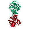
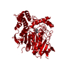
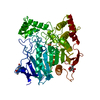
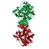
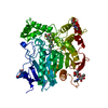
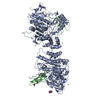
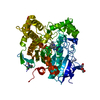
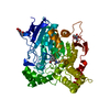
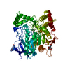
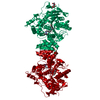
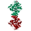
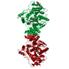
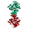
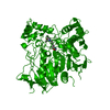
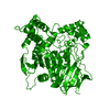
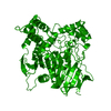
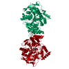
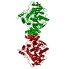
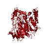
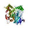
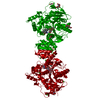
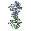
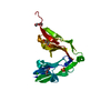
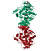
| Title | Structure of acetylcholinesterase (E.C. 3.1.1.7) complexed with N-9-(1',2',3',4'-Tetrahydroacridinyl)-1,8- diaminooctane at 2.4 angstroms resolution. | ||||||
 Components Components | ACETYLCHOLINESTERASE | ||||||
 Keywords Keywords | HYDROLASE / SERINE HYDROLASE / ACETYLCHOLINESTERASE / NEUROTRANSMITTER CLEAVAGE / ALZHEIMER'S DISEASE / BIVALENT LIGAND / DUAL-SITE BINDING / INHIBITOR / TACRINE / AMINE / HETERODIMER / SERINE ESTERASE SYNAPSE / MEMBRANE / NERVE / MUSCLE / GPI-ANCHOR NEUROTRANSMITTER DEGRADATION / GLYCOPROTEIN | ||||||
| Function / homology |  Function and homology information Function and homology informationacetylcholine catabolic process in synaptic cleft / acetylcholinesterase / choline metabolic process / acetylcholinesterase activity / side of membrane / synaptic cleft / synapse / : / plasma membrane Similarity search - Function | ||||||
| Biological species |   TORPEDO CALIFORNICA (Pacific electric ray) TORPEDO CALIFORNICA (Pacific electric ray) | ||||||
| Method |  X-RAY DIFFRACTION / MOLECULAR REPLACEMENT / Resolution: 2.4 Å X-RAY DIFFRACTION / MOLECULAR REPLACEMENT / Resolution: 2.4 Å | ||||||
 Authors Authors | Brumshtein, B. / Wong, D.M. / Greenblatt, H.M. / Carlier, P.R. / Pang, Y.-P. / Silman, I. / Sussman, J.L. | ||||||
 Citation Citation | Journal: J.Med.Chem. / Year: 2006 Title: Complexes of Alkylene-Linked Tacrine Dimers with Torpedo Californica Acetylcholinesterase: Binding of Bis(5)-Tacrine Produces a Dramatic Rearrangement in the Active-Site Gorge. Authors: Rydberg, E.H. / Brumshtein, B. / Greenblatt, H.M. / Wong, D.M. / Shaya, D. / Williams, L.D. / Carlier, P.R. / Pang, Y.-P. / Silman, I. / Sussman, J.L. #1: Journal: J.Am.Chem.Soc. / Year: 2003Title: Acetylcholinesterase Complexed with Bivalent Ligands Related to Huperzine A: Experimental Evidence for Species-Dependent Protein-Ligand Complementarity Authors: Wong, D.M. / Greenblatt, H.M. / Dvir, H. / Carlier, P.R. / Han, Y.-F. / Pang, Y.-P. / Silman, I. / Sussman, J.L. #2: Journal: J.Med.Chem. / Year: 1999 Title: Heterodimeric Tacrine-Based Acetylcholinesterase Inhibitors: Investigating Ligand-Peripheral Site Interactions Authors: Carlier, P.R. / Chow, E.S.-H. / Han, Y.-F. / Liu, J. / El Yazal, J. / Pang, Y.-P. #3: Journal: J. Comput. Aided Mol. Des. / Year: 1994 Title: Prediction of the Binding Sites of Huperzine a in Acetylcholinesterase by Docking Studies Authors: Pang, Y.-P. / Kozikowski, A.P. #4: Journal: Proc.Natl.Acad.Sci.USA / Year: 1993Title: Quaternary Ligand Binding to Aromatic Residues in the Active-Site Gorge of Acetylcholinesterase Authors: Harel, M. / Schalk, I. / Ehret-Sabatier, L. / Bouet, F. / Goeldner, M. / Hirth, C. / Axelsen, P.H. / Silman, I. / Sussman, J.L. #5: Journal: Science / Year: 1991 Title: Atomic Structure of Acetylcholinesterase from Torpedo Californica: A Prototypic Acetylcholine-Binding Protein Authors: Sussman, J.L. / Harel, M. / Frolow, F. / Oefner, C. / Goldman, A. / Toker, L. / Silman, I. | ||||||
| History |
|
- Structure visualization
| Structure viewer | Molecule: MolmilJmol/JSmol |
|---|
- Downloads & links
-Download
| PDBx/mmCIF format | 1ut6.cif.gz | 123.6 KB | Display | PDBx/mmCIF format |
|---|---|---|---|---|
| PDB format | pdb1ut6.ent.gz | 95.4 KB | Display | PDB format |
| PDBx/mmJSON format | 1ut6.json.gz | Tree view | PDBx/mmJSON format | |
| Others |  Other downloads Other downloads |
-Validation report
| Arichive directory | https://data.pdbj.org/pub/pdb/validation_reports/ut/1ut6ftp://data.pdbj.org/pub/pdb/validation_reports/ut/1ut6 | HTTPS FTP |
|---|
-Related structure data
| Related structure data |  1odcC  2ckmC  2cmfC  2aceS C: citing same article ( S: Starting model for refinement |
|---|---|
| Similar structure data |


-Links
 PDBj
PDBj
- Assembly
| Deposited unit | 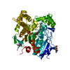
| ||||||||
|---|---|---|---|---|---|---|---|---|---|
| 1 | 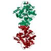
| ||||||||
| Unit cell |
|
-Components
| #1: Protein | Mass: 60792.516 Da / Num. of mol.: 1 / Source method: isolated from a natural source Details: SYNTHETIC HETEROBIVALENT TACRINE-BASED DIMER, A8NH2 (N-9-(1', 2', 3', 4'-TETRAHYDROACRIDINYL)-1, 8-DIAMINOOCTANE) DIHYDROCHLORIDE WITH THE TACRINE MOIETY BOUND TO THE 'ANIONIC' SUBSITE, NEAR ...Details: SYNTHETIC HETEROBIVALENT TACRINE-BASED DIMER, A8NH2 (N-9-(1', 2', 3', 4'-TETRAHYDROACRIDINYL)-1, 8-DIAMINOOCTANE) DIHYDROCHLORIDE WITH THE TACRINE MOIETY BOUND TO THE 'ANIONIC' SUBSITE, NEAR THE BOTTOM OF THE ACTIVE SITE GORGE, AND THE 8-AMINO MOIETY BOUND TO THE 'PERIPHERAL' ANIONIC SITE (PAS) AT THE TOP OF THE GORGE OF TCACHE. THE LIGAND, A8NH2, THUS BINDS BY SPANNING THE ACTIVE-SITE GORGE. Source: (natural) TORPEDO CALIFORNICA (Pacific electric ray)Organ: ELECTRIC ORGAN / Variant: G2 FORM / Tissue: ELECTROPLAQUE / References: UniProt: P04058, acetylcholinesterase | ||||||||||
|---|---|---|---|---|---|---|---|---|---|---|---|
| #2: Sugar |   Type: D-saccharide, beta linking / Mass: 221.208 Da / Num. of mol.: 2 Type: D-saccharide, beta linking / Mass: 221.208 Da / Num. of mol.: 2Source method: isolated from a genetically manipulated source Formula: C8H15NO6 #3: Chemical | ChemComp-A8N / |   Mass: 327.507 Da / Num. of mol.: 1 / Source method: obtained synthetically / Formula: C21H33N3 Mass: 327.507 Da / Num. of mol.: 1 / Source method: obtained synthetically / Formula: C21H33N3#4: Water | ChemComp-HOH / |  Mass: 18.015 Da / Num. of mol.: 160 / Source method: isolated from a natural source / Formula: H2O Mass: 18.015 Da / Num. of mol.: 160 / Source method: isolated from a natural source / Formula: H2OCompound details | PROTEIN HYDROLYZES | Has protein modification | Y | Sequence details | THE SEQUENCE DESCRIBED BELOW IS THAT OF THE T-ISOFORM SPLICE VARIANT OF SWISSPROT ENTRY P04058 AND ...THE SEQUENCE DESCRIBED BELOW IS THAT OF THE T-ISOFORM SPLICE VARIANT OF SWISSPROT ENTRY P04058 AND ANNOTATED IN FTID=VSP_001460 GLU A 536 SWS P04058 ALA 557 THR A 537 SWS P04058 CYS 558 | |
-Experimental details
-Experiment
| Experiment | Method: X-RAY DIFFRACTION / Number of used crystals: 1 |
|---|
- Sample preparation
| Crystal | Density Matthews: 4.04 Å3/Da / Density % sol: 69.56 % |
|---|---|
| Crystal grow | Temperature: 277 K / pH: 5.8 Details: PROTEIN WAS CRYSTALLISED FROM 28-33% V/V PEG 200 0.5M MES PH 5.8 AT 4 DEG. CELSIUS; THEN SOAKED IN MOTHER LIQUOR (40% V/V PEG 200 IN 0.1 M MES BUFFER, PH 5.8) CONTAINING 2MM N-9-(1',2',3',4'- ...Details: PROTEIN WAS CRYSTALLISED FROM 28-33% V/V PEG 200 0.5M MES PH 5.8 AT 4 DEG. CELSIUS; THEN SOAKED IN MOTHER LIQUOR (40% V/V PEG 200 IN 0.1 M MES BUFFER, PH 5.8) CONTAINING 2MM N-9-(1',2',3',4'-TETRAHYDROACRIDINYL) -1,8-DIAMINOOCTANE BIS-HYDROCHLORIDE (A8NH2.2HCL) |
-Data collection
| Diffraction | Mean temperature: 120 K |
|---|---|
| Diffraction source | Source: ROTATING ANODE / Type: RIGAKU RUH3R / Wavelength: 1.5418 |
| Detector | Type: RIGAKU IMAGE PLATE / Detector: IMAGE PLATE / Date: Jan 18, 2001 / Details: OSMIC BLUE CONFOCAL MIRRO |
| Radiation | Protocol: SINGLE WAVELENGTH / Monochromatic (M) / Laue (L): M / Scattering type: x-ray |
| Radiation wavelength | Wavelength: 1.5418 Å / Relative weight: 1 |
| Reflection | Resolution: 2.4→30 Å / Num. obs: 38785 / % possible obs: 97 % / Redundancy: 3.22 % / Biso Wilson estimate: 37.2 Å2 / Rmerge(I) obs: 0.053 / Net I/σ(I): 16.9 |
| Reflection shell | Resolution: 2.4→2.49 Å / Redundancy: 9.95 % / Rmerge(I) obs: 0.397 / Mean I/σ(I) obs: 2.44 / % possible all: 97.5 |
- Processing
| Software |
| ||||||||||||||||||||||||||||||||||||||||||||||||||||||||||||||||||||||||||||||||
|---|---|---|---|---|---|---|---|---|---|---|---|---|---|---|---|---|---|---|---|---|---|---|---|---|---|---|---|---|---|---|---|---|---|---|---|---|---|---|---|---|---|---|---|---|---|---|---|---|---|---|---|---|---|---|---|---|---|---|---|---|---|---|---|---|---|---|---|---|---|---|---|---|---|---|---|---|---|---|---|---|---|
| Refinement | Method to determine structure: MOLECULAR REPLACEMENT Starting model: PDB ENTRY 2ACE Resolution: 2.4→29.14 Å / Rfactor Rfree error: 0.005 / Data cutoff high absF: 1843636.65 / Isotropic thermal model: RESTRAINED / Cross valid method: THROUGHOUT / σ(F): 0 Details: SEVERAL RESIDUES ARE NOT SEEN IN THE CRYSTAL STRUCTURE, DUE TO DISORDER. THESE INCLUDE ASP A1, ASP A2, HIS A3 AND THE C-TERMINAL RESIDUES AFTER THR A535. SEVERAL RESIDUES MISSING IN CHAIN ...Details: SEVERAL RESIDUES ARE NOT SEEN IN THE CRYSTAL STRUCTURE, DUE TO DISORDER. THESE INCLUDE ASP A1, ASP A2, HIS A3 AND THE C-TERMINAL RESIDUES AFTER THR A535. SEVERAL RESIDUES MISSING IN CHAIN BREAK, FROM HIS A486 - GLU A489 (INCLUSIVE).
| ||||||||||||||||||||||||||||||||||||||||||||||||||||||||||||||||||||||||||||||||
| Solvent computation | Solvent model: FLAT MODEL / Bsol: 38.7999 Å2 / ksol: 0.351354 e/Å3 | ||||||||||||||||||||||||||||||||||||||||||||||||||||||||||||||||||||||||||||||||
| Displacement parameters | Biso mean: 46.7 Å2
| ||||||||||||||||||||||||||||||||||||||||||||||||||||||||||||||||||||||||||||||||
| Refine analyze |
| ||||||||||||||||||||||||||||||||||||||||||||||||||||||||||||||||||||||||||||||||
| Refinement step | Cycle: LAST / Resolution: 2.4→29.14 Å
| ||||||||||||||||||||||||||||||||||||||||||||||||||||||||||||||||||||||||||||||||
| Refine LS restraints |
| ||||||||||||||||||||||||||||||||||||||||||||||||||||||||||||||||||||||||||||||||
| LS refinement shell | Resolution: 2.4→2.49 Å / Rfactor Rfree error: 0.024 / Total num. of bins used: 10
| ||||||||||||||||||||||||||||||||||||||||||||||||||||||||||||||||||||||||||||||||
| Xplor file |
|
