 Movie
Movie Controller

[English] 日本語
 Yorodumi
- PDB-2uu8: X-ray structure of Ni, Ca concanavalin A at Ultra-high resolution... -
+ Open data

- Basic information
| Entry | Database: PDB / ID: 2uu8 | ||||||
|---|---|---|---|---|---|---|---|
| Title | X-ray structure of Ni, Ca concanavalin A at Ultra-high resolution (0. 94A) | ||||||
 Components Components | CONCANAVALIN | ||||||
 Keywords Keywords | LECTIN / CALCIUM / MANGANESE / METAL-BINDING / NI | ||||||
| Function / homology |  Function and homology information Function and homology informationregulation of defense response to virus / D-mannose binding / defense response / metal ion binding Similarity search - Function | ||||||
| Biological species |   CANAVALIA ENSIFORMIS (jack bean) CANAVALIA ENSIFORMIS (jack bean) | ||||||
| Method |  X-RAY DIFFRACTION / SYNCHROTRON / MOLECULAR REPLACEMENT / Resolution: 0.94 Å X-RAY DIFFRACTION / SYNCHROTRON / MOLECULAR REPLACEMENT / Resolution: 0.94 Å | ||||||
 Authors Authors | Ahmed, H.U. / Blakeley, M.P. / Cianci, M. / Cruickshank, D.W.J. / Hubbard, J.A. / Helliwell, J.R. | ||||||
 Citation Citation | Journal: Acta Crystallogr.,Sect.D / Year: 2007 Title: The Determination of Protonation States in Proteins. Authors: Ahmed, H.U. / Blakeley, M.P. / Cianci, M. / Cruickshank, D.W.J. / Hubbard, J.A. / Helliwell, J.R. | ||||||
| History |
| ||||||
| Remark 700 | SHEET THE SHEET STRUCTURE OF THIS MOLECULE IS BIFURCATED. IN ORDER TO REPRESENT THIS FEATURE IN ... SHEET THE SHEET STRUCTURE OF THIS MOLECULE IS BIFURCATED. IN ORDER TO REPRESENT THIS FEATURE IN THE SHEET RECORDS BELOW, TWO SHEETS ARE DEFINED. |
- Structure visualization
| Structure viewer | Molecule: MolmilJmol/JSmol |
|---|
- Downloads & links
-Download
| PDBx/mmCIF format | 2uu8.cif.gz | 121.3 KB | Display | PDBx/mmCIF format |
|---|---|---|---|---|
| PDB format | pdb2uu8.ent.gz | 92.8 KB | Display | PDB format |
| PDBx/mmJSON format | 2uu8.json.gz | Tree view | PDBx/mmJSON format | |
| Others |  Other downloads Other downloads |
-Validation report
| Arichive directory | https://data.pdbj.org/pub/pdb/validation_reports/uu/2uu8ftp://data.pdbj.org/pub/pdb/validation_reports/uu/2uu8 | HTTPS FTP |
|---|
-Related structure data
| Related structure data |  2uufC  2uujC 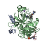 2uukC  2yz4C  1nlsS S: Starting model for refinement C: citing same article ( |
|---|---|
| Similar structure data |


-Links
 PDBj
PDBj

- Assembly
| Deposited unit | 
| ||||||||
|---|---|---|---|---|---|---|---|---|---|
| 1 | 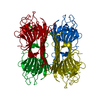
| ||||||||
| Unit cell |
|
-Components
| #1: Protein | Mass: 25622.385 Da / Num. of mol.: 1 / Source method: isolated from a natural source / Source: (natural) CANAVALIA ENSIFORMIS (jack bean) / References: UniProt: P02866 |
|---|---|
| #2: Chemical | ChemComp-NI /   Mass: 58.693 Da / Num. of mol.: 1 / Source method: obtained synthetically / Formula: Ni Mass: 58.693 Da / Num. of mol.: 1 / Source method: obtained synthetically / Formula: Ni |
| #3: Chemical | ChemComp-CA /   Mass: 40.078 Da / Num. of mol.: 1 / Source method: obtained synthetically / Formula: Ca Mass: 40.078 Da / Num. of mol.: 1 / Source method: obtained synthetically / Formula: Ca |
| #4: Water | ChemComp-HOH /  Mass: 18.015 Da / Num. of mol.: 287 / Source method: isolated from a natural source / Formula: H2O Mass: 18.015 Da / Num. of mol.: 287 / Source method: isolated from a natural source / Formula: H2O |
| Sequence details | THE MATURE CHAIN IS FORMED THROUGH A POSTTRANSLATIONAL MODIFICATION AFTER THE REMOVAL OF THE SIGNAL ...THE MATURE CHAIN IS FORMED THROUGH A POSTTRANSL |
-Experimental details
-Experiment
| Experiment | Method: X-RAY DIFFRACTION / Number of used crystals: 3 |
|---|
- Sample preparation
| Crystal | Density Matthews: 1.98 Å3/Da / Density % sol: 37.8 % / Description: NONE |
|---|---|
| Crystal grow | pH: 6.5 / Details: pH 6.5 |
-Data collection
| Diffraction | Mean temperature: 100 K |
|---|---|
| Diffraction source | Source: SYNCHROTRON / Site: APS  / Beamline: 19-ID / Wavelength: 0.67 / Beamline: 19-ID / Wavelength: 0.67 |
| Detector | Type: ADSC CCD / Detector: CCD / Date: Oct 15, 2004 |
| Radiation | Monochromator: SI(III) / Protocol: SINGLE WAVELENGTH / Monochromatic (M) / Laue (L): M / Scattering type: x-ray |
| Radiation wavelength | Wavelength: 0.67 Å / Relative weight: 1 |
| Reflection | Resolution: 0.94→33.23 Å / Num. obs: 136027 / % possible obs: 89.2 % / Redundancy: 6 % / Rmerge(I) obs: 0.09 / Net I/σ(I): 11.84 |
| Reflection shell | Resolution: 0.94→0.99 Å / Redundancy: 2.5 % / Rmerge(I) obs: 0.33 / Mean I/σ(I) obs: 1.98 / % possible all: 54.4 |
- Processing
| Software |
| |||||||||||||||||||||||||||||||||
|---|---|---|---|---|---|---|---|---|---|---|---|---|---|---|---|---|---|---|---|---|---|---|---|---|---|---|---|---|---|---|---|---|---|---|
| Refinement | Method to determine structure: MOLECULAR REPLACEMENT Starting model: PDB ENTRY 1NLS Resolution: 0.94→33.23 Å / Num. parameters: 19691 / Num. restraintsaints: 24823 / Cross valid method: FREE R-VALUE
| |||||||||||||||||||||||||||||||||
| Refine analyze | Num. disordered residues: 37 / Occupancy sum hydrogen: 1752.31 / Occupancy sum non hydrogen: 2084.35 | |||||||||||||||||||||||||||||||||
| Refinement step | Cycle: LAST / Resolution: 0.94→33.23 Å
| |||||||||||||||||||||||||||||||||
| Refine LS restraints |
|
