 Movie
Movie Controller


+ Open data

- Basic information
| Entry | Database: PDB / ID: 1joj | ||||||
|---|---|---|---|---|---|---|---|
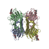
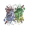
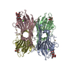
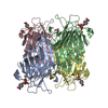
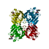
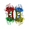
| Title | CONCANAVALIN A-HEXAPEPTIDE COMPLEX | ||||||
 Components Components |
| ||||||
 Keywords Keywords | SUGAR BINDING PROTEIN / lectin | ||||||
| Function / homology |  Function and homology information Function and homology information | ||||||
| Biological species |   Canavalia ensiformis (jack bean) Canavalia ensiformis (jack bean)synthetic construct (others) | ||||||
| Method |  X-RAY DIFFRACTION / MOLECULAR REPLACEMENT / Resolution: 3 Å X-RAY DIFFRACTION / MOLECULAR REPLACEMENT / Resolution: 3 Å | ||||||
 Authors Authors | Jain, D. / Kaur, K. / Salunke, D.M. | ||||||
 Citation Citation | Journal: Biochemistry / Year: 2001 Title: Enhanced binding of a rationally designed peptide ligand of concanavalin a arises from improved geometrical complementarity. Authors: Jain, D. / Kaur, K.J. / Salunke, D.M. #1: Journal: Biophys.J. / Year: 2001Title: Plasticity in Protein-Peptide Recognition: Crystal Structures of Two Different Peptides Bound to Concanavalin A Authors: Jain, D. / Kaur, K.J. / Salunke, D.M. #2: Journal: J.Biol.Chem. / Year: 2000Title: Structural and Functional Consequences of Peptide-Carbohydrate Mimicry: Crystal Structure of a Carbohydrate-Mimicking Peptide Bound to Concanavalin A Authors: Jain, D. / Kaur, K.J. / Sundaravadivel, B. / Salunke, D.M. | ||||||
| History |
|
- Structure visualization
| Structure viewer | Molecule: MolmilJmol/JSmol |
|---|
- Downloads & links
-Download
| PDBx/mmCIF format | 1joj.cif.gz | 184.6 KB | Display | PDBx/mmCIF format |
|---|---|---|---|---|
| PDB format | pdb1joj.ent.gz | 150 KB | Display | PDB format |
| PDBx/mmJSON format | 1joj.json.gz | Tree view | PDBx/mmJSON format | |
| Others |  Other downloads Other downloads |
-Validation report
| Arichive directory | https://data.pdbj.org/pub/pdb/validation_reports/jo/1jojftp://data.pdbj.org/pub/pdb/validation_reports/jo/1joj | HTTPS FTP |
|---|
-Related structure data
| Related structure data |  5cnaS S: Starting model for refinement |
|---|---|
| Similar structure data |


-Links
 PDBj
PDBj

- Assembly
| Deposited unit | 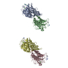
| ||||||||
|---|---|---|---|---|---|---|---|---|---|
| 1 | 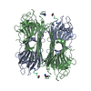
| ||||||||
| 2 | 
| ||||||||
| Unit cell |
|
-Components
| #1: Protein | Mass: 25622.385 Da / Num. of mol.: 4 / Source method: isolated from a natural source / Source: (natural) Canavalia ensiformis (jack bean) / References: UniProt: P55915#2: Protein/peptide | Mass: 946.103 Da / Num. of mol.: 4 / Source method: obtained synthetically / Details: The peptide was chemically synthesized. / Source: (synth.) synthetic construct (others) #3: Chemical | ChemComp-MN /   Mass: 54.938 Da / Num. of mol.: 4 / Source method: obtained synthetically / Formula: Mn Mass: 54.938 Da / Num. of mol.: 4 / Source method: obtained synthetically / Formula: Mn#4: Chemical | ChemComp-CA /   Mass: 40.078 Da / Num. of mol.: 4 / Source method: obtained synthetically / Formula: Ca Mass: 40.078 Da / Num. of mol.: 4 / Source method: obtained synthetically / Formula: Ca#5: Water | ChemComp-HOH / |  Mass: 18.015 Da / Num. of mol.: 197 / Source method: isolated from a natural source / Formula: H2O Mass: 18.015 Da / Num. of mol.: 197 / Source method: isolated from a natural source / Formula: H2OHas protein modification | Y | |
|---|
-Experimental details
-Experiment
| Experiment | Method: X-RAY DIFFRACTION |
|---|
- Sample preparation
| Crystal | Density Matthews: 3.62 Å3/Da / Density % sol: 66.06 % | ||||||||||||||||||||||||||||||
|---|---|---|---|---|---|---|---|---|---|---|---|---|---|---|---|---|---|---|---|---|---|---|---|---|---|---|---|---|---|---|---|
| Crystal grow | *PLUS pH: 9 / Method: vapor diffusion, hanging drop | ||||||||||||||||||||||||||||||
| Components of the solutions | *PLUS
|
-Data collection
| Diffraction source | Source: ROTATING ANODE / Type: RIGAKU RU200 / Wavelength: 1.54178 |
|---|---|
| Detector | Detector: IMAGE PLATE |
| Radiation | Protocol: SINGLE WAVELENGTH / Monochromatic (M) / Laue (L): M / Scattering type: x-ray |
| Radiation wavelength | Wavelength: 1.54178 Å / Relative weight: 1 |
| Reflection | Highest resolution: 3 Å / Num. obs: 23957 / % possible obs: 78.1 % / Redundancy: 3 % / Biso Wilson estimate: 24.3 Å2 / Rmerge(I) obs: 0.101 / Net I/σ(I): 10.6 |
| Reflection shell | Resolution: 3→3.13 Å / % possible obs: 71.8 % / Mean I/σ(I) obs: 5.37 / % possible all: 71.8 |
| Reflection shell | *PLUS Rmerge(I) obs: 0.257 |
- Processing
| Software |
| ||||||||||||||||||||||||||||||||||||||||||||||||||||||||||||||||||||||||||||||||
|---|---|---|---|---|---|---|---|---|---|---|---|---|---|---|---|---|---|---|---|---|---|---|---|---|---|---|---|---|---|---|---|---|---|---|---|---|---|---|---|---|---|---|---|---|---|---|---|---|---|---|---|---|---|---|---|---|---|---|---|---|---|---|---|---|---|---|---|---|---|---|---|---|---|---|---|---|---|---|---|---|---|
| Refinement | Method to determine structure: MOLECULAR REPLACEMENT Starting model: 5CNA Resolution: 3→10 Å / Cross valid method: THROUGHOUT / σ(F): 0
| ||||||||||||||||||||||||||||||||||||||||||||||||||||||||||||||||||||||||||||||||
| Displacement parameters | Biso mean: 28.1 Å2 | ||||||||||||||||||||||||||||||||||||||||||||||||||||||||||||||||||||||||||||||||
| Refine analyze |
| ||||||||||||||||||||||||||||||||||||||||||||||||||||||||||||||||||||||||||||||||
| Refinement step | Cycle: LAST / Resolution: 3→10 Å
| ||||||||||||||||||||||||||||||||||||||||||||||||||||||||||||||||||||||||||||||||
| Refine LS restraints |
| ||||||||||||||||||||||||||||||||||||||||||||||||||||||||||||||||||||||||||||||||
| Xplor file |
| ||||||||||||||||||||||||||||||||||||||||||||||||||||||||||||||||||||||||||||||||
| Software | *PLUS Name: CNS / Version: 0.5 / Classification: refinement | ||||||||||||||||||||||||||||||||||||||||||||||||||||||||||||||||||||||||||||||||
| Refinement | *PLUS σ(F): 0 / % reflection Rfree: 4.8 % / Rfactor obs: 0.186 | ||||||||||||||||||||||||||||||||||||||||||||||||||||||||||||||||||||||||||||||||
| Solvent computation | *PLUS | ||||||||||||||||||||||||||||||||||||||||||||||||||||||||||||||||||||||||||||||||
| Displacement parameters | *PLUS Biso mean: 28.1 Å2 | ||||||||||||||||||||||||||||||||||||||||||||||||||||||||||||||||||||||||||||||||
| Refine LS restraints | *PLUS
|
