 Movie
Movie Controller

[English] 日本語
 Yorodumi
- PDB-3d4k: Concanavalin A Complexed to a Synthetic Analog of the Trimannoside -
+ Open data

- Basic information
| Entry | Database: PDB / ID: 3d4k | |||||||||
|---|---|---|---|---|---|---|---|---|---|---|
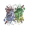
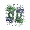
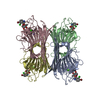
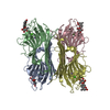
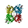
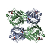
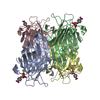
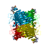
| Title | Concanavalin A Complexed to a Synthetic Analog of the Trimannoside | |||||||||
 Components Components | Concanavalin-A | |||||||||
 Keywords Keywords | SUGAR BINDING PROTEIN / Concanavalin A / Conserved Water / Carbohydrate-Protein Binding / Glycoprotein / Lectin / Manganese / Metal-binding | |||||||||
| Function / homology |  Function and homology information Function and homology informationregulation of defense response to virus / D-mannose binding / defense response / metal ion binding Similarity search - Function | |||||||||
| Biological species |   Canavalia ensiformis (jack bean) Canavalia ensiformis (jack bean) | |||||||||
| Method |  X-RAY DIFFRACTION / SYNCHROTRON / MOLECULAR REPLACEMENT / molecular replacement / Resolution: 1.8 Å X-RAY DIFFRACTION / SYNCHROTRON / MOLECULAR REPLACEMENT / molecular replacement / Resolution: 1.8 Å | |||||||||
 Authors Authors | Kadirvelraj, R. / Foley, B.L. / Dyekjaer, J.D. / Woods, R.J. | |||||||||
 Citation Citation | Journal: J.Am.Chem.Soc. / Year: 2008 Title: Involvement of water in carbohydrate-protein binding: concanavalin A revisited. Authors: Kadirvelraj, R. / Foley, B.L. / Dyekjaer, J.D. / Woods, R.J. #1: Journal: J.Am.Chem.Soc. / Year: 2001 Title: Involvement of water in carbohydrate-protein binding. Authors: Clarke, C. / Woods, R.J. / Gluska, J. / Cooper, A. / Nutley, M.A. / Boons, G.J. #2: Journal: J.Biol.Chem. / Year: 1996Title: Structural basis of trimannoside recognition by concanavalin A. Authors: Naismith, J.H. / Field, R.A. | |||||||||
| History |
|
- Structure visualization
| Structure viewer | Molecule: MolmilJmol/JSmol |
|---|
- Downloads & links
-Download
| PDBx/mmCIF format | 3d4k.cif.gz | 208.7 KB | Display | PDBx/mmCIF format |
|---|---|---|---|---|
| PDB format | pdb3d4k.ent.gz | 165.9 KB | Display | PDB format |
| PDBx/mmJSON format | 3d4k.json.gz | Tree view | PDBx/mmJSON format | |
| Others |  Other downloads Other downloads |
-Validation report
| Arichive directory | https://data.pdbj.org/pub/pdb/validation_reports/d4/3d4kftp://data.pdbj.org/pub/pdb/validation_reports/d4/3d4k | HTTPS FTP |
|---|
-Related structure data
| Related structure data |  1qd0S S: Starting model for refinement |
|---|---|
| Similar structure data |


-Links
 PDBj
PDBj

- Assembly
| Deposited unit | 
| ||||||||
|---|---|---|---|---|---|---|---|---|---|
| 1 | 
| ||||||||
| Unit cell |
|
-Components
-Protein / Sugars , 2 types, 8 molecules ABCD
| #1: Protein | Mass: 25622.385 Da / Num. of mol.: 4 / Source method: isolated from a natural source / Details: Jack-bean / Source: (natural) Canavalia ensiformis (jack bean) / References: UniProt: P02866#2: Polysaccharide | alpha-D-mannopyranose-(1-3)-[alpha-D-mannopyranose-(1-6)]methyl 2-deoxy-2-(2-hydroxyethyl)-alpha-D- ...alpha-D-mannopyranose-(1-3)-[alpha-D-mannopyranose-(1-6)]methyl 2-deoxy-2-(2-hydroxyethyl)-alpha-D-mannopyranoside Type: oligosaccharide / Mass: 546.518 Da / Num. of mol.: 4 / Source method: obtained synthetically |
|---|
-Non-polymers , 4 types, 584 molecules 


| #3: Chemical | ChemComp-MN /  Mass: 54.938 Da / Num. of mol.: 4 / Source method: obtained synthetically / Formula: Mn Mass: 54.938 Da / Num. of mol.: 4 / Source method: obtained synthetically / Formula: Mn#4: Chemical | ChemComp-CA /  Mass: 40.078 Da / Num. of mol.: 4 / Source method: obtained synthetically / Formula: Ca Mass: 40.078 Da / Num. of mol.: 4 / Source method: obtained synthetically / Formula: Ca#5: Chemical | ChemComp-GOL /  Mass: 92.094 Da / Num. of mol.: 4 / Source method: obtained synthetically / Formula: C3H8O3 Mass: 92.094 Da / Num. of mol.: 4 / Source method: obtained synthetically / Formula: C3H8O3#6: Water | ChemComp-HOH / | Mass: 18.015 Da / Num. of mol.: 572 / Source method: isolated from a natural source / Formula: H2O |
|---|
-Experimental details
-Experiment
| Experiment | Method: X-RAY DIFFRACTION / Number of used crystals: 1 |
|---|
- Sample preparation
| Crystal | Density Matthews: 2.34 Å3/Da / Density % sol: 47.55 % |
|---|---|
| Crystal grow | Temperature: 291 K / Method: vapor diffusion, hanging drop / pH: 6.8 Details: 10-16% PEG 6000, 100 mM Sodium cacodylate, 50 mM NaCL, 1 mM MnCL2, 1 mM CaCL2, pH 6.8, VAPOR DIFFUSION, HANGING DROP, temperature 291K |
-Data collection
| Diffraction | Mean temperature: 100 K |
|---|---|
| Diffraction source | Source: SYNCHROTRON / Site: CHESS  / Beamline: A1 / Wavelength: 0.935 Å / Beamline: A1 / Wavelength: 0.935 Å |
| Detector | Type: ADSC QUANTUM 210 / Detector: CCD / Date: Mar 14, 2002 / Details: Mirrors |
| Radiation | Monochromator: Si (111) / Protocol: SINGLE WAVELENGTH / Scattering type: x-ray |
| Radiation wavelength | Wavelength: 0.935 Å / Relative weight: 1 |
| Reflection | Resolution: 1.8→100 Å / Num. all: 88130 / Num. obs: 87296 / % possible obs: 99.1 % / Observed criterion σ(F): 1 / Observed criterion σ(I): 1 / Redundancy: 4.1 % / Biso Wilson estimate: 20.3 Å2 / Rmerge(I) obs: 0.06 / Rsym value: 0.036 / Χ2: 1.057 / Net I/σ(I): 13.5 |
| Reflection shell | Resolution: 1.8→1.86 Å / Redundancy: 1.01 % / Rmerge(I) obs: 0.566 / Mean I/σ(I) obs: 2.9 / Num. unique all: 8649 / Rsym value: 0.502 / Χ2: 1.063 / % possible all: 98.7 |
-Phasing
| Phasing | Method: molecular replacement | |||||||||
|---|---|---|---|---|---|---|---|---|---|---|
| Phasing MR | Rfactor: 0.576 / Cor.coef. Fo:Fc: 0.13 / Cor.coef. Io to Ic: 0.12
|
- Processing
| Software |
| ||||||||||||||||||||||||||||
|---|---|---|---|---|---|---|---|---|---|---|---|---|---|---|---|---|---|---|---|---|---|---|---|---|---|---|---|---|---|
| Refinement | Method to determine structure: MOLECULAR REPLACEMENT Starting model: pdb entry 1QD0 Resolution: 1.8→30 Å / FOM work R set: 0.83 / Isotropic thermal model: Isotropic / Cross valid method: SigmaA / σ(F): 0 / σ(I): 0 / Stereochemistry target values: Engh & Huber
| ||||||||||||||||||||||||||||
| Solvent computation | Bsol: 52.702 Å2 | ||||||||||||||||||||||||||||
| Displacement parameters | Biso mean: 24.552 Å2
| ||||||||||||||||||||||||||||
| Refine analyze | Luzzati coordinate error obs: 0.223 Å / Luzzati d res low obs: 30 Å / Luzzati sigma a obs: 0.076 Å | ||||||||||||||||||||||||||||
| Refinement step | Cycle: LAST / Resolution: 1.8→30 Å
| ||||||||||||||||||||||||||||
| Refine LS restraints |
| ||||||||||||||||||||||||||||
| LS refinement shell | Resolution: 1.8→1.86 Å
| ||||||||||||||||||||||||||||
| Xplor file |
|
