 Movie
Movie Controller

[English] 日本語
 Yorodumi
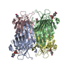
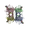
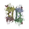
- PDB-1cjp: CONCANAVALIN A COMPLEX WITH 4'-METHYLUMBELLIFERYL-ALPHA-D-GLUCOPY... -
+ Open data

- Basic information
| Entry | Database: PDB / ID: 1cjp | ||||||
|---|---|---|---|---|---|---|---|
| Title | CONCANAVALIN A COMPLEX WITH 4'-METHYLUMBELLIFERYL-ALPHA-D-GLUCOPYRANOSIDE | ||||||
 Components Components | CONCANAVALIN A | ||||||
 Keywords Keywords | LECTIN / LEGUME LECTIN | ||||||
| Function / homology |  Function and homology information Function and homology informationregulation of defense response to virus / D-mannose binding / defense response / metal ion binding Similarity search - Function | ||||||
| Biological species |   Canavalia ensiformis (jack bean) Canavalia ensiformis (jack bean) | ||||||
| Method |  X-RAY DIFFRACTION / MOLECULAR REPLACEMENT / Resolution: 2.78 Å X-RAY DIFFRACTION / MOLECULAR REPLACEMENT / Resolution: 2.78 Å | ||||||
 Authors Authors | Hamodrakas, S.J. / Kanellopoulos, P.N. / Tucker, P.A. | ||||||
 Citation Citation | Journal: J.Struct.Biol. / Year: 1997 Title: The crystal structure of the complex of concanavalin A with 4'-methylumbelliferyl-alpha-D-glucopyranoside. Authors: Hamodrakas, S.J. / Kanellopoulos, P.N. / Pavlou, K. / Tucker, P.A. #1: Journal: J.Struct.Biol. / Year: 1996Title: A Triclinic Crystal Form of the Lectin Concanavalin A Authors: Kanellopoulos, P.N. / Tucker, P.A. / Pavlou, K. / Agianian, B. / Hamodrakas, S.J. #2: Journal: J.Struct.Biol. / Year: 1996Title: The Crystal Structure of the Complexes of Concanavalin a with 4'-Nitrophenyl-Alpha-D-Mannopyranoside and 4'-Nitrophenyl-Alpha-D-Glucopyranoside Authors: Kanellopoulos, P.N. / Pavlou, K. / Perrakis, A. / Agianian, B. / Vorgias, C.E. / Mavrommatis, C. / Soufi, M. / Tucker, P.A. / Hamodrakas, S.J. | ||||||
| History |
|
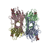
- Structure visualization
| Structure viewer | Molecule: MolmilJmol/JSmol |
|---|
- Downloads & links
-Download
| PDBx/mmCIF format | 1cjp.cif.gz | 193.2 KB | Display | PDBx/mmCIF format |
|---|---|---|---|---|
| PDB format | pdb1cjp.ent.gz | 154.3 KB | Display | PDB format |
| PDBx/mmJSON format | 1cjp.json.gz | Tree view | PDBx/mmJSON format | |
| Others |  Other downloads Other downloads |
-Validation report
| Arichive directory | https://data.pdbj.org/pub/pdb/validation_reports/cj/1cjpftp://data.pdbj.org/pub/pdb/validation_reports/cj/1cjp | HTTPS FTP |
|---|
-Related structure data
| Related structure data |  1vamS S: Starting model for refinement |
|---|---|
| Similar structure data |


-Links
 PDBj
PDBj

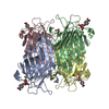
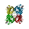
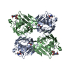
- Assembly
| Deposited unit | 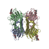
| ||||||||
|---|---|---|---|---|---|---|---|---|---|
| 1 |
| ||||||||
| Unit cell |
|
-Components
| #1: Protein | Mass: 25622.385 Da / Num. of mol.: 4 / Source method: isolated from a natural source / Source: (natural) Canavalia ensiformis (jack bean) / References: UniProt: P02866#2: Sugar | ChemComp-MUG /   Type: D-saccharide / Mass: 338.309 Da / Num. of mol.: 4 Type: D-saccharide / Mass: 338.309 Da / Num. of mol.: 4Source method: isolated from a genetically manipulated source Formula: C16H18O8 #3: Chemical | ChemComp-MN /   Mass: 54.938 Da / Num. of mol.: 4 / Source method: obtained synthetically / Formula: Mn Mass: 54.938 Da / Num. of mol.: 4 / Source method: obtained synthetically / Formula: Mn#4: Chemical | ChemComp-CA /   Mass: 40.078 Da / Num. of mol.: 4 / Source method: obtained synthetically / Formula: Ca Mass: 40.078 Da / Num. of mol.: 4 / Source method: obtained synthetically / Formula: Ca#5: Water | ChemComp-HOH / |  Mass: 18.015 Da / Num. of mol.: 162 / Source method: isolated from a natural source / Formula: H2O Mass: 18.015 Da / Num. of mol.: 162 / Source method: isolated from a natural source / Formula: H2O |
|---|
-Experimental details
-Experiment
| Experiment | Method: X-RAY DIFFRACTION |
|---|
- Sample preparation
| Crystal | Density Matthews: 3.7 Å3/Da / Density % sol: 66.78 % | ||||||||||||||||||||
|---|---|---|---|---|---|---|---|---|---|---|---|---|---|---|---|---|---|---|---|---|---|
| Crystal grow | *PLUS pH: 8.5 / Method: vapor diffusion, hanging drop | ||||||||||||||||||||
| Components of the solutions | *PLUS
|
-Data collection
| Diffraction source | Wavelength: 1.5418 |
|---|---|
| Detector | Type: MARRESEARCH / Detector: IMAGE PLATE / Date: Nov 30, 1994 |
| Radiation | Monochromatic (M) / Laue (L): M / Scattering type: x-ray |
| Radiation wavelength | Wavelength: 1.5418 Å / Relative weight: 1 |
| Reflection | Num. obs: 37358 / % possible obs: 99 % / Observed criterion σ(I): 0 / Redundancy: 4 % / Rmerge(I) obs: 0.074 |
| Reflection | *PLUS Highest resolution: 2.78 Å / Lowest resolution: 48 Å / Num. measured all: 151116 |
| Reflection shell | *PLUS Highest resolution: 2.78 Å / Lowest resolution: 2.9 Å / % possible obs: 95.2 % / Rmerge(I) obs: 0.238 |
- Processing
| Software |
| ||||||||||||||||||||||||||||||||||||||||||||||||||||||||||||
|---|---|---|---|---|---|---|---|---|---|---|---|---|---|---|---|---|---|---|---|---|---|---|---|---|---|---|---|---|---|---|---|---|---|---|---|---|---|---|---|---|---|---|---|---|---|---|---|---|---|---|---|---|---|---|---|---|---|---|---|---|---|
| Refinement | Method to determine structure: MOLECULAR REPLACEMENT Starting model: PDB ENTRY 1VAM Resolution: 2.78→8 Å / σ(F): 0
| ||||||||||||||||||||||||||||||||||||||||||||||||||||||||||||
| Displacement parameters | Biso mean: 51.41 Å2 | ||||||||||||||||||||||||||||||||||||||||||||||||||||||||||||
| Refine analyze | Luzzati coordinate error obs: 0.25 Å | ||||||||||||||||||||||||||||||||||||||||||||||||||||||||||||
| Refinement step | Cycle: LAST / Resolution: 2.78→8 Å
| ||||||||||||||||||||||||||||||||||||||||||||||||||||||||||||
| Refine LS restraints |
|
