| 登録情報 | データベース: PDB / ID: 1jn2
|
|---|
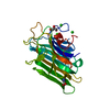
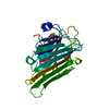
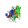
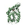
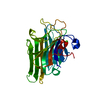
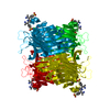
| タイトル | Crystal Structure of meso-tetrasulphonatophenyl porphyrin complexed with Concanavalin A |
|---|
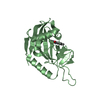
 要素 要素 | Concanavalin-A |
|---|
 キーワード キーワード | SUGAR BINDING PROTEIN / LECTIN |
|---|
| 機能・相同性 |  機能・相同性情報 機能・相同性情報
regulation of defense response to virus / D-mannose binding / defense response / metal ion binding類似検索 - 分子機能 Legume lectin / Legume lectin, alpha chain, conserved site / Legume lectins alpha-chain signature. / Legume lectins beta-chain signature. / Legume lectin domain / Legume lectin domain / : / Legume lectin, beta chain, Mn/Ca-binding site / Jelly Rolls - #200 / Concanavalin A-like lectin/glucanase domain superfamily ...Legume lectin / Legume lectin, alpha chain, conserved site / Legume lectins alpha-chain signature. / Legume lectins beta-chain signature. / Legume lectin domain / Legume lectin domain / : / Legume lectin, beta chain, Mn/Ca-binding site / Jelly Rolls - #200 / Concanavalin A-like lectin/glucanase domain superfamily / Jelly Rolls / Sandwich / Mainly Beta類似検索 - ドメイン・相同性 |
|---|
| 生物種 |   Canavalia ensiformis (タチナタマメ) Canavalia ensiformis (タチナタマメ) |
|---|
| 手法 |  X線回折 / 分子置換 / 解像度: 1.9 Å X線回折 / 分子置換 / 解像度: 1.9 Å |
|---|
 データ登録者 データ登録者 | Goel, M. / Jain, D. / Kaur, K.J. / Kenoth, R. / Maiya, B.G. / Swamy, M.J. / Salunke, D.M. |
|---|
 引用 引用 | ジャーナル: J.Biol.Chem. / 年: 2000
タイトル: Functional equality in the absence of structural similarity: an added dimension to molecular mimicry
著者: Goel, M. / Jain, D. / Kaur, K.J. / Kenoth, R. / Maiya, B.G. / Swamy, M.J. / Salunke, D.M. |
|---|
| 履歴 | | 登録 | 2001年7月22日 | 登録サイト: RCSB / 処理サイト: RCSB |
|---|
| 改定 1.0 | 2003年7月1日 | Provider: repository / タイプ: Initial release |
|---|
| 改定 1.1 | 2008年4月27日 | Group: Version format compliance |
|---|
| 改定 1.2 | 2011年7月13日 | Group: Derived calculations / Version format compliance |
|---|
| 改定 1.3 | 2017年6月28日 | Group: Database references / Source and taxonomy / Structure summary
カテゴリ: entity / entity_name_com ...entity / entity_name_com / entity_src_nat / struct / struct_ref / struct_ref_seq / struct_ref_seq_dif
Item: _entity.pdbx_description / _entity.pdbx_fragment ..._entity.pdbx_description / _entity.pdbx_fragment / _struct.pdbx_descriptor / _struct_ref_seq.ref_id |
|---|
| 改定 1.4 | 2024年2月7日 | Group: Data collection / Database references / Derived calculations
カテゴリ: chem_comp_atom / chem_comp_bond ...chem_comp_atom / chem_comp_bond / database_2 / struct_conn / struct_site
Item: _database_2.pdbx_DOI / _database_2.pdbx_database_accession ..._database_2.pdbx_DOI / _database_2.pdbx_database_accession / _struct_conn.pdbx_dist_value / _struct_conn.ptnr1_label_atom_id / _struct_conn.ptnr2_auth_comp_id / _struct_conn.ptnr2_auth_seq_id / _struct_conn.ptnr2_label_asym_id / _struct_conn.ptnr2_label_atom_id / _struct_conn.ptnr2_label_comp_id / _struct_site.pdbx_auth_asym_id / _struct_site.pdbx_auth_comp_id / _struct_site.pdbx_auth_seq_id |
|---|
|
|---|
| Remark 999 | SEQUENCE THE MODEL USED WAS 5CNA WHICH HAS THE SAME SEQUENCE AS THIS STRUCTURE. |
|---|
 ムービー
ムービー コントローラー


 データを開く
 基本情報
基本情報 構造の表示
構造の表示 ダウンロードとリンク
 その他のダウンロード


 PDBj
PDBj

 集合体
集合体

 分子量: 40.078 Da / 分子数: 1 / 由来タイプ: 合成 / 式: Ca
 分子量: 54.938 Da / 分子数: 1 / 由来タイプ: 合成 / 式: Mn
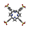
 分子量: 939.020 Da / 分子数: 1 / 由来タイプ: 合成 / 式: C44H34N4O12S4
 分子量: 18.015 Da / 分子数: 51 / 由来タイプ: 天然 / 式: H2O
 試料調製
試料調製 解析
解析