 Movie
Movie Controller

[English] 日本語
 Yorodumi
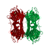
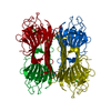
- PDB-1apn: THE CRYSTALLOGRAPHIC STRUCTURE OF METAL-FREE CONCANAVALIN A AT 2.... -
+ Open data

- Basic information
| Entry | Database: PDB / ID: 1apn | ||||||
|---|---|---|---|---|---|---|---|
| Title | THE CRYSTALLOGRAPHIC STRUCTURE OF METAL-FREE CONCANAVALIN A AT 2.5 ANGSTROMS RESOLUTION | ||||||
 Components Components | CONCANAVALIN A | ||||||
 Keywords Keywords | LECTIN | ||||||
| Function / homology |  Function and homology information Function and homology informationregulation of defense response to virus / D-mannose binding / defense response / metal ion binding Similarity search - Function | ||||||
| Biological species |   Canavalia ensiformis (jack bean) Canavalia ensiformis (jack bean) | ||||||
| Method |  X-RAY DIFFRACTION / Resolution: 2.5 Å X-RAY DIFFRACTION / Resolution: 2.5 Å | ||||||
 Authors Authors | Bouckaert, J. / Loris, R. / Poortmans, F. / Wyns, L. | ||||||
 Citation Citation | Journal: Proteins / Year: 1995 Title: Crystallographic structure of metal-free concanavalin A at 2.5 A resolution. Authors: Bouckaert, J. / Loris, R. / Poortmans, F. / Wyns, L. #1: Journal: J.Mol.Biol. / Year: 1979Title: Crystal Structure of Demetallized Concanavalin A. The Metal-Binding Region Authors: Shoham, M. / Yonath, A. / Sussman, J.L. / Moult, J. / Traub, W. / Gilboa(Kalb), A.J. #2: Journal: Proc.Natl.Acad.Sci.USA / Year: 1978Title: Changes in the Three-Dimensional Structure of Concanavalin a Upon Demetallization Authors: Reeke Junior, G.N. / Becker, J.W. / Edelman, G.M. | ||||||
| History |
|
- Structure visualization
| Structure viewer | Molecule: MolmilJmol/JSmol |
|---|
- Downloads & links
-Download
| PDBx/mmCIF format | 1apn.cif.gz | 99.6 KB | Display | PDBx/mmCIF format |
|---|---|---|---|---|
| PDB format | pdb1apn.ent.gz | 77.3 KB | Display | PDB format |
| PDBx/mmJSON format | 1apn.json.gz | Tree view | PDBx/mmJSON format | |
| Others |  Other downloads Other downloads |
-Validation report
| Arichive directory | https://data.pdbj.org/pub/pdb/validation_reports/ap/1apnftp://data.pdbj.org/pub/pdb/validation_reports/ap/1apn | HTTPS FTP |
|---|
-Related structure data
| Similar structure data |
|---|


-Links
 PDBj
PDBj

- Assembly
| Deposited unit | 
| ||||||||
|---|---|---|---|---|---|---|---|---|---|
| 1 | 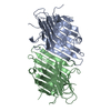
| ||||||||
| Unit cell |
| ||||||||
| Atom site foot note | 1: GLU A 122 - THR A 123 OMEGA = 210.44 PEPTIDE BOND DEVIATES SIGNIFICANTLY FROM TRANS CONFORMATION | ||||||||
| Noncrystallographic symmetry (NCS) | NCS oper: (Code: given Matrix: (0.970545, 0.240787, 0.007974), Vector: Details | MTRIX THE TRANSFORMATIONS PRESENTED ON MTRIX RECORDS BELOW DESCRIBE NON-CRYSTALLOGRAPHIC RELATIONSHIPS AMONG THE VARIOUS DOMAINS IN THIS ENTRY. APPLYING THE APPROPRIATE MTRIX TRANSFORMATION TO THE RESIDUES LISTED FIRST WILL YIELD APPROXIMATE COORDINATES FOR THE RESIDUES LISTED SECOND. APPLIED TO TRANSFORMED TO MTRIX RESIDUES RESIDUES RMSD M1 B 1 .. B 237 A 1 .. A 237 0.559 | |
-Components
| #1: Protein | Mass: 25622.385 Da / Num. of mol.: 2 / Source method: isolated from a natural source / Details: DEMETALLIZED, PH 5 / Source: (natural) Canavalia ensiformis (jack bean) / References: UniProt: P02866#2: Water | ChemComp-HOH / |  Mass: 18.015 Da / Num. of mol.: 168 / Source method: isolated from a natural source / Formula: H2O Mass: 18.015 Da / Num. of mol.: 168 / Source method: isolated from a natural source / Formula: H2O |
|---|
-Experimental details
-Experiment
| Experiment | Method: X-RAY DIFFRACTION |
|---|
- Sample preparation
| Crystal | Density Matthews: 2.34 Å3/Da / Density % sol: 47.41 % | ||||||||||||||||||||||||||||||||||||
|---|---|---|---|---|---|---|---|---|---|---|---|---|---|---|---|---|---|---|---|---|---|---|---|---|---|---|---|---|---|---|---|---|---|---|---|---|---|
| Crystal | *PLUS Density % sol: 44 % | ||||||||||||||||||||||||||||||||||||
| Crystal grow | *PLUS pH: 5 / Method: vapor diffusion, sitting drop | ||||||||||||||||||||||||||||||||||||
| Components of the solutions | *PLUS
|
-Data collection
| Diffraction source | Wavelength: 1.5418 |
|---|---|
| Detector | Detector: AREA DETECTOR / Date: Jan 20, 1993 |
| Radiation | Monochromatic (M) / Laue (L): M / Scattering type: x-ray |
| Radiation wavelength | Wavelength: 1.5418 Å / Relative weight: 1 |
| Reflection | Resolution: 2.5→15 Å / Num. obs: 16834 / % possible obs: 98 % / Observed criterion σ(I): 0 / Rmerge(I) obs: 0.078 |
| Reflection | *PLUS Num. measured all: 88305 / Rmerge(I) obs: 0.078 |
- Processing
| Software |
| ||||||||||||||||||||||||||||||||||||||||||||||||||||||||||||
|---|---|---|---|---|---|---|---|---|---|---|---|---|---|---|---|---|---|---|---|---|---|---|---|---|---|---|---|---|---|---|---|---|---|---|---|---|---|---|---|---|---|---|---|---|---|---|---|---|---|---|---|---|---|---|---|---|---|---|---|---|---|
| Refinement | Resolution: 2.5→10 Å / σ(F): 0
| ||||||||||||||||||||||||||||||||||||||||||||||||||||||||||||
| Displacement parameters | Biso mean: 32 Å2 | ||||||||||||||||||||||||||||||||||||||||||||||||||||||||||||
| Refinement step | Cycle: LAST / Resolution: 2.5→10 Å
| ||||||||||||||||||||||||||||||||||||||||||||||||||||||||||||
| Refine LS restraints |
| ||||||||||||||||||||||||||||||||||||||||||||||||||||||||||||
| Software | *PLUS Name: X-PLOR / Classification: refinement | ||||||||||||||||||||||||||||||||||||||||||||||||||||||||||||
| Refinement | *PLUS | ||||||||||||||||||||||||||||||||||||||||||||||||||||||||||||
| Solvent computation | *PLUS | ||||||||||||||||||||||||||||||||||||||||||||||||||||||||||||
| Displacement parameters | *PLUS | ||||||||||||||||||||||||||||||||||||||||||||||||||||||||||||
| Refine LS restraints | *PLUS
|
