 Movie
Movie Controller

[English] 日本語
 Yorodumi
- PDB-1con: THE REFINED STRUCTURE OF CADMIUM SUBSTITUTED CONCANAVALIN A AT 2.... -
+ Open data

- Basic information
| Entry | Database: PDB / ID: 1con | ||||||
|---|---|---|---|---|---|---|---|
| Title | THE REFINED STRUCTURE OF CADMIUM SUBSTITUTED CONCANAVALIN A AT 2.0 ANGSTROMS RESOLUTION | ||||||
 Components Components | CONCANAVALIN A | ||||||
 Keywords Keywords | LECTIN(AGGLUTININ) | ||||||
| Function / homology |  Function and homology information Function and homology informationregulation of defense response to virus / D-mannose binding / defense response / metal ion binding Similarity search - Function | ||||||
| Biological species |   Canavalia ensiformis (jack bean) Canavalia ensiformis (jack bean) | ||||||
| Method |  X-RAY DIFFRACTION / Resolution: 2 Å X-RAY DIFFRACTION / Resolution: 2 Å | ||||||
 Authors Authors | Naismith, J.H. / Habash, J. / Harrop, S.J. / Helliwell, J.R. / Hunter, W.N. / Kalb(Gilboa), A.J. / Yariv, J. / Wan, T.C.M. / Weisgerber, S. | ||||||
 Citation Citation | Journal: Acta Crystallogr.,Sect.D / Year: 1993 Title: Refined structure of cadmium-substituted concanavalin A at 2.0 A resolution. Authors: Naismith, J.H. / Habash, J. / Harrop, S. / Helliwell, J.R. / Hunter, W.N. / Wan, T.C. / Weisgerber, S. / Kalb, A.J. / Yariv, J. #1: Journal: Embo J. / Year: 1992Title: Non-Glycosylated Recombinant Pro-Concanavalin a is Active without Polypeptide Cleavage Authors: Min, W. / Dunn, A.J. / Jones, D.H. | ||||||
| History |
| ||||||
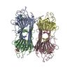
| Remark 700 | SHEET THE MOST STRIKING FEATURE OF CONCANAVALIN A IS TWO LARGE BETA SHEETS IN THE STRUCTURE ...SHEET THE MOST STRIKING FEATURE OF CONCANAVALIN A IS TWO LARGE BETA SHEETS IN THE STRUCTURE COMPRISING 111 OF THE 237 AMINO ACIDS. THERE IS NO ALPHA HELIX. THE REMAINING AMINO ACIDS FORM A SERIES OF LOOPS AND TURNS. THE PURPOSE OF THIS STUDY IS TO PROVIDE A WELL-REFINED SACCHARIDE-FREE STRUCTURE BASED UPON THE CORRECTED AMINO ACID SEQUENCE. THIS STRUCTURE IS PART OF A BROADER STUDY TO DEFINE THE PROTEIN SACCHARIDE INTERACTIONS OF CONCANAVALIN A. |
- Structure visualization
| Structure viewer | Molecule: MolmilJmol/JSmol |
|---|
- Downloads & links
-Download
| PDBx/mmCIF format | 1con.cif.gz | 61.6 KB | Display | PDBx/mmCIF format |
|---|---|---|---|---|
| PDB format | pdb1con.ent.gz | 45 KB | Display | PDB format |
| PDBx/mmJSON format | 1con.json.gz | Tree view | PDBx/mmJSON format | |
| Others |  Other downloads Other downloads |
-Validation report
| Arichive directory | https://data.pdbj.org/pub/pdb/validation_reports/co/1conftp://data.pdbj.org/pub/pdb/validation_reports/co/1con | HTTPS FTP |
|---|
-Related structure data
| Similar structure data |
|---|


-Links
 PDBj
PDBj

- Assembly
| Deposited unit | 
| ||||||||
|---|---|---|---|---|---|---|---|---|---|
| 1 | 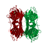
| ||||||||
| Unit cell |
| ||||||||
| Atom site foot note | 1: ALA A 207 - ASP A 208 OMEGA = 0.16 PEPTIDE BOND DEVIATES SIGNIFICANTLY FROM TRANS CONFORMATION 2: THE FOLLOWING SURFACE LOOPS ARE IN WEAK DENSITY: A 118 - A 123, A 149 - A 152, AND A 159 - A 162. | ||||||||
| Components on special symmetry positions |
|
-Components
| #1: Protein | Mass: 25622.385 Da / Num. of mol.: 1 Source method: isolated from a genetically manipulated source Source: (gene. exp.) Canavalia ensiformis (jack bean) / Organ: BEAN / References: UniProt: P02866 | ||||||
|---|---|---|---|---|---|---|---|
| #2: Chemical |   Mass: 112.411 Da / Num. of mol.: 2 / Source method: obtained synthetically / Formula: Cd Mass: 112.411 Da / Num. of mol.: 2 / Source method: obtained synthetically / Formula: Cd#3: Chemical | ChemComp-CA / |   Mass: 40.078 Da / Num. of mol.: 1 / Source method: obtained synthetically / Formula: Ca Mass: 40.078 Da / Num. of mol.: 1 / Source method: obtained synthetically / Formula: Ca#4: Water | ChemComp-HOH / |  Mass: 18.015 Da / Num. of mol.: 144 / Source method: isolated from a natural source / Formula: H2O Mass: 18.015 Da / Num. of mol.: 144 / Source method: isolated from a natural source / Formula: H2OSequence details | THE AUTHORS STATE THAT THIS IS THE CORRECT AMINO ACID SEQUENCE FOR CONCANAVALIN A, ACCORDING TO THE ...THE AUTHORS STATE THAT THIS IS THE CORRECT AMINO ACID SEQUENCE FOR CONCANAVAL | |
-Experimental details
-Experiment
| Experiment | Method: X-RAY DIFFRACTION |
|---|
- Sample preparation
| Crystal | Density Matthews: 2.34 Å3/Da / Density % sol: 47.4 % |
|---|---|
| Crystal grow | *PLUS Method: other / Details: Kalb, A.J., (1988) J. Crystal Growth, 88, 537. |
-Data collection
| Radiation | Scattering type: x-ray |
|---|---|
| Radiation wavelength | Relative weight: 1 |
| Reflection | *PLUS Highest resolution: 2 Å / Lowest resolution: 8 Å / Num. obs: 16178 / % possible obs: 99.1 % / Rmerge(I) obs: 0.06 |
| Reflection shell | *PLUS Highest resolution: 2 Å / Lowest resolution: 2.1 Å / Num. unique obs: 2188 / Rmerge(I) obs: 0.113 |
- Processing
| Software |
| ||||||||||||||||||||||||||||||||||||||||||||||||||||||||||||
|---|---|---|---|---|---|---|---|---|---|---|---|---|---|---|---|---|---|---|---|---|---|---|---|---|---|---|---|---|---|---|---|---|---|---|---|---|---|---|---|---|---|---|---|---|---|---|---|---|---|---|---|---|---|---|---|---|---|---|---|---|---|
| Refinement | Resolution: 2→8 Å / σ(F): 0 /
| ||||||||||||||||||||||||||||||||||||||||||||||||||||||||||||
| Refinement step | Cycle: LAST / Resolution: 2→8 Å
| ||||||||||||||||||||||||||||||||||||||||||||||||||||||||||||
| Refine LS restraints |
| ||||||||||||||||||||||||||||||||||||||||||||||||||||||||||||
| Refinement | *PLUS Highest resolution: 2 Å / Lowest resolution: 8 Å / Num. reflection all: 16178 / σ(F): 0 / Rfactor all: 0.171 | ||||||||||||||||||||||||||||||||||||||||||||||||||||||||||||
| Solvent computation | *PLUS | ||||||||||||||||||||||||||||||||||||||||||||||||||||||||||||
| Displacement parameters | *PLUS | ||||||||||||||||||||||||||||||||||||||||||||||||||||||||||||
| Refine LS restraints | *PLUS
|
