 Movie
Movie Controller

[English] 日本語
 Yorodumi
- PDB-1ces: CRYSTALS OF DEMETALLIZED CONCANAVALIN A SOAKED WITH ZINC HAVE A Z... -
+ Open data

- Basic information
| Entry | Database: PDB / ID: 1ces | ||||||
|---|---|---|---|---|---|---|---|
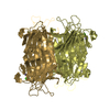
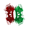
| Title | CRYSTALS OF DEMETALLIZED CONCANAVALIN A SOAKED WITH ZINC HAVE A ZINC ION BOUND IN THE S1 SITE | ||||||
 Components Components | CONCANAVALIN A | ||||||
 Keywords Keywords | LECTIN / CONCANAVALIN A / ZINC / CONFORMATION | ||||||
| Function / homology |  Function and homology information Function and homology informationregulation of defense response to virus / D-mannose binding / defense response / metal ion binding Similarity search - Function | ||||||
| Biological species |   Canavalia ensiformis (jack bean) Canavalia ensiformis (jack bean) | ||||||
| Method |  X-RAY DIFFRACTION / MOLECULAR REPLACEMENT / Resolution: 2.7 Å X-RAY DIFFRACTION / MOLECULAR REPLACEMENT / Resolution: 2.7 Å | ||||||
 Authors Authors | Bouckaert, J. / Loris, R. / Poortmans, F. / Wyns, L. | ||||||
 Citation Citation | Journal: J.Biol.Chem. / Year: 1996 Title: Sequential structural changes upon zinc and calcium binding to metal-free concanavalin A. Authors: Bouckaert, J. / Poortmans, F. / Wyns, L. / Loris, R. #1: Journal: Proteins / Year: 1995Title: Crystallographic Structure of Metal-Free Concanavalin a at 2.5 A Resolution Authors: Bouckaert, J. / Loris, R. / Poortmans, F. / Wyns, L. | ||||||
| History |
| ||||||
| Remark 700 | SHEET THE MOST STRIKING FEATURE OF CONCANAVALIN A IS TWO LARGE BETA SHEETS IN THE STRUCTURE ...SHEET THE MOST STRIKING FEATURE OF CONCANAVALIN A IS TWO LARGE BETA SHEETS IN THE STRUCTURE COMPRISING 119 OF THE 237 AMINO ACIDS. THERE IS NO ALPHA HELIX. THE REMAINING AMINO ACIDS FORM A SERIES OF LOOPS AND TURNS. CONCANAVALIN A EXISTS AS A TETRAMER ALTHOUGH THE ASYMMETRIC UNIT CONTAINS A DIMER. THE TETRAMER CAN BE GENERATED BY SYMMETRY OPERATORS. |
- Structure visualization
| Structure viewer | Molecule: MolmilJmol/JSmol |
|---|
- Downloads & links
-Download
| PDBx/mmCIF format | 1ces.cif.gz | 97.6 KB | Display | PDBx/mmCIF format |
|---|---|---|---|---|
| PDB format | pdb1ces.ent.gz | 74.6 KB | Display | PDB format |
| PDBx/mmJSON format | 1ces.json.gz | Tree view | PDBx/mmJSON format | |
| Others |  Other downloads Other downloads |
-Validation report
| Arichive directory | https://data.pdbj.org/pub/pdb/validation_reports/ce/1cesftp://data.pdbj.org/pub/pdb/validation_reports/ce/1ces | HTTPS FTP |
|---|
-Related structure data
| Related structure data |  1enqC  1enrC  1ensC  1apnS S: Starting model for refinement C: citing same article ( |
|---|---|
| Similar structure data |


-Links
 PDBj
PDBj

- Assembly
| Deposited unit | 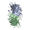
| ||||||||
|---|---|---|---|---|---|---|---|---|---|
| 1 | 
| ||||||||
| Unit cell |
|
-Components
| #1: Protein | Mass: 25622.385 Da / Num. of mol.: 2 / Source method: isolated from a natural source Details: THERE ARE TWO MONOMERS (MR 25500 EACH) WITH IDENTICAL SEQUENCE Source: (natural) Canavalia ensiformis (jack bean) / References: UniProt: P02866#2: Chemical |   Mass: 65.409 Da / Num. of mol.: 2 / Source method: obtained synthetically / Formula: Zn Mass: 65.409 Da / Num. of mol.: 2 / Source method: obtained synthetically / Formula: Zn#3: Water | ChemComp-HOH / |  Mass: 18.015 Da / Num. of mol.: 39 / Source method: isolated from a natural source / Formula: H2O Mass: 18.015 Da / Num. of mol.: 39 / Source method: isolated from a natural source / Formula: H2ONonpolymer details | ZINC ION IS PARTIALLY BOUND IN THE S1 SITE. | |
|---|
-Experimental details
-Experiment
| Experiment | Method: X-RAY DIFFRACTION / Number of used crystals: 1 |
|---|
- Sample preparation
| Crystal | Density Matthews: 2.35 Å3/Da / Density % sol: 45.9 % | ||||||||||||||||||||||||||||||||||||||||
|---|---|---|---|---|---|---|---|---|---|---|---|---|---|---|---|---|---|---|---|---|---|---|---|---|---|---|---|---|---|---|---|---|---|---|---|---|---|---|---|---|---|
| Crystal grow | pH: 5 / Details: pH 5. | ||||||||||||||||||||||||||||||||||||||||
| Crystal grow | *PLUS Method: vapor diffusion, sitting drop | ||||||||||||||||||||||||||||||||||||||||
| Components of the solutions | *PLUS
|
-Data collection
| Diffraction | Mean temperature: 291 K |
|---|---|
| Diffraction source | Source: ROTATING ANODE / Type: ENRAF-NONIUS / Wavelength: 1.5418 |
| Detector | Type: ENRAF-NONIUS FAST / Detector: DIFFRACTOMETER / Date: Jun 9, 1993 |
| Radiation | Monochromatic (M) / Laue (L): M / Scattering type: x-ray |
| Radiation wavelength | Wavelength: 1.5418 Å / Relative weight: 1 |
| Reflection | Resolution: 2.7→10 Å / Num. obs: 12557 / % possible obs: 91.5 % / Observed criterion σ(I): 0 / Rmerge(I) obs: 0.085 |
| Reflection | *PLUS Num. measured all: 29303 |
- Processing
| Software |
| ||||||||||||||||||||||||||||||||||||||||||||||||||||||||||||
|---|---|---|---|---|---|---|---|---|---|---|---|---|---|---|---|---|---|---|---|---|---|---|---|---|---|---|---|---|---|---|---|---|---|---|---|---|---|---|---|---|---|---|---|---|---|---|---|---|---|---|---|---|---|---|---|---|---|---|---|---|---|
| Refinement | Method to determine structure: MOLECULAR REPLACEMENT Starting model: DEMETALLIZED CONCANAVALIN A (PDB ENTRY 1APN) Resolution: 2.7→10 Å / Cross valid method: BRUNGER / σ(F): 0
| ||||||||||||||||||||||||||||||||||||||||||||||||||||||||||||
| Displacement parameters | Biso mean: 24.89 Å2 | ||||||||||||||||||||||||||||||||||||||||||||||||||||||||||||
| Refine analyze | Luzzati coordinate error obs: 0.25 Å | ||||||||||||||||||||||||||||||||||||||||||||||||||||||||||||
| Refinement step | Cycle: LAST / Resolution: 2.7→10 Å
| ||||||||||||||||||||||||||||||||||||||||||||||||||||||||||||
| Refine LS restraints |
| ||||||||||||||||||||||||||||||||||||||||||||||||||||||||||||
| Software | *PLUS Name: X-PLOR / Classification: refinement | ||||||||||||||||||||||||||||||||||||||||||||||||||||||||||||
| Refinement | *PLUS | ||||||||||||||||||||||||||||||||||||||||||||||||||||||||||||
| Solvent computation | *PLUS | ||||||||||||||||||||||||||||||||||||||||||||||||||||||||||||
| Displacement parameters | *PLUS | ||||||||||||||||||||||||||||||||||||||||||||||||||||||||||||
| Refine LS restraints | *PLUS
|
