 Movie
Movie Controller

[English] 日本語
 Yorodumi
- PDB-1enq: CO-CRYSTALS OF DEMETALLIZED CONCANAVALIN A WITH ZINC HAVING A ZIN... -
+ Open data

- Basic information
| Entry | Database: PDB / ID: 1enq | ||||||
|---|---|---|---|---|---|---|---|
| Title | CO-CRYSTALS OF DEMETALLIZED CONCANAVALIN A WITH ZINC HAVING A ZINC ION BOUND IN THE S1 SITE | ||||||
 Components Components | CONCANAVALIN A | ||||||
 Keywords Keywords | PLANT LECTIN (AGGLUTININ) / CONCANAVALIN A / ZINC / PLANT LECTIN / AGGLUTININ | ||||||
| Function / homology |  Function and homology information Function and homology informationregulation of defense response to virus / D-mannose binding / defense response / metal ion binding Similarity search - Function | ||||||
| Biological species |   Canavalia ensiformis (jack bean) Canavalia ensiformis (jack bean) | ||||||
| Method |  X-RAY DIFFRACTION / Resolution: 2.5 Å X-RAY DIFFRACTION / Resolution: 2.5 Å | ||||||
 Authors Authors | Bouckaert, J. / Loris, R. / Poortmans, F. / Wyns, L. | ||||||
 Citation Citation | Journal: J.Biol.Chem. / Year: 1996 Title: Sequential structural changes upon zinc and calcium binding to metal-free concanavalin A. Authors: Bouckaert, J. / Poortmans, F. / Wyns, L. / Loris, R. #1: Journal: Proteins / Year: 1995Title: Crystallographic Structure of Metal-Free Concanavalin a at 2.5 Angstrom Resolution Authors: Bouckaert, J. / Loris, R. / Poortmans, F. / Wyns, L. | ||||||
| History |
| ||||||
| Remark 700 | SHEET THE MOST STRIKING FEATURE OF CONCANAVALIN A IS TWO LARGE BETA SHEETS IN THE STRUCTURE ...SHEET THE MOST STRIKING FEATURE OF CONCANAVALIN A IS TWO LARGE BETA SHEETS IN THE STRUCTURE COMPRISING 119 OF THE 237 AMINO ACIDS. THERE IS NO ALPHA HELIX. THE REMAINING AMINO ACIDS FORM A SERIES OF LOOPS AND TURNS. |
- Structure visualization
| Structure viewer | Molecule: MolmilJmol/JSmol |
|---|
- Downloads & links
-Download
| PDBx/mmCIF format | 1enq.cif.gz | 189.6 KB | Display | PDBx/mmCIF format |
|---|---|---|---|---|
| PDB format | pdb1enq.ent.gz | 152.9 KB | Display | PDB format |
| PDBx/mmJSON format | 1enq.json.gz | Tree view | PDBx/mmJSON format | |
| Others |  Other downloads Other downloads |
-Validation report
| Arichive directory | https://data.pdbj.org/pub/pdb/validation_reports/en/1enqftp://data.pdbj.org/pub/pdb/validation_reports/en/1enq | HTTPS FTP |
|---|
-Related structure data


-Links
 PDBj
PDBj

- Assembly
| Deposited unit | 
| ||||||||
|---|---|---|---|---|---|---|---|---|---|
| 1 |
| ||||||||
| Unit cell |
| ||||||||
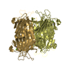
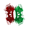
| Details | THE ASYMMETRIC UNIT CONTAINS FOUR MONOMERS EACH CONTAINING 237 AMINO ACIDS (CHAINS A, B, C, AND D), FORMING A BIOLOGICALLY FUNCTIONAL TETRAMER. THE FOLLOWING RESIDUES ARE IN VERY WEAK DENSITY IN ALL FOUR MONOMERS: 99 - 101, 117 - 122, 160 - 167, 204 - 204, 235 - 237. THE SURFACE LOOP RESIDUES 14 - 19 ARE IN WEAK DENSITY IN CHAIN B, ARE BADLY DEFINED BY THE ELECTRON DENSITY IN CHAIN C, AND UNDEFINED IN CHAIN D. THE ATOMS WERE MODELLED AS OXT OF THE PREVIOUS RESIDUE. NO DENSITY IS OBSERVED FOR THE REST OF THE RESIDUE. PDB CHOSE REPRESENT THESE ATOMS AS THE N ATOM OF THE PROCEEDING RESIDU N SER A 161 N SER B 160 N SER C 160 N THR D 15 N SER D 160 |
-Components
| #1: Protein | Mass: 25622.385 Da / Num. of mol.: 4 / Source method: isolated from a natural source / Details: ZINC COMPLEX / Source: (natural) Canavalia ensiformis (jack bean) / References: UniProt: P02866#2: Chemical | ChemComp-ZN /   Mass: 65.409 Da / Num. of mol.: 4 / Source method: obtained synthetically / Formula: Zn Mass: 65.409 Da / Num. of mol.: 4 / Source method: obtained synthetically / Formula: Zn#3: Water | ChemComp-HOH / |  Mass: 18.015 Da / Num. of mol.: 295 / Source method: isolated from a natural source / Formula: H2O Mass: 18.015 Da / Num. of mol.: 295 / Source method: isolated from a natural source / Formula: H2O |
|---|
-Experimental details
-Experiment
| Experiment | Method: X-RAY DIFFRACTION |
|---|
- Sample preparation
| Crystal | Density Matthews: 2.26 Å3/Da / Density % sol: 47.4 % | ||||||||||||||||||||||||||||||||||||||||||||||||||||||
|---|---|---|---|---|---|---|---|---|---|---|---|---|---|---|---|---|---|---|---|---|---|---|---|---|---|---|---|---|---|---|---|---|---|---|---|---|---|---|---|---|---|---|---|---|---|---|---|---|---|---|---|---|---|---|---|
| Crystal grow | pH: 5 / Details: pH 5. | ||||||||||||||||||||||||||||||||||||||||||||||||||||||
| Crystal | *PLUS Density % sol: 43.7 % | ||||||||||||||||||||||||||||||||||||||||||||||||||||||
| Crystal grow | *PLUS pH: 5 / Method: vapor diffusion, sitting drop | ||||||||||||||||||||||||||||||||||||||||||||||||||||||
| Components of the solutions | *PLUS
|
-Data collection
| Diffraction source | Wavelength: 1.5418 |
|---|---|
| Detector | Type: ENRAF-NONIUS FAST / Detector: DIFFRACTOMETER / Date: Jan 30, 1995 |
| Radiation | Monochromatic (M) / Laue (L): M / Scattering type: x-ray |
| Radiation wavelength | Wavelength: 1.5418 Å / Relative weight: 1 |
| Reflection | Resolution: 2.19→42 Å / Num. obs: 32457 / % possible obs: 97.8 % / Observed criterion σ(I): 0 / Rmerge(I) obs: 0.011 |
| Reflection | *PLUS Highest resolution: 2.49 Å / Lowest resolution: 15 Å / Num. obs: 31981 / % possible obs: 97.5 % / Num. measured all: 121921 / Rmerge(I) obs: 0.0112 |
- Processing
| Software |
| ||||||||||||||||||||||||||||||||||||||||||||||||||||||||||||
|---|---|---|---|---|---|---|---|---|---|---|---|---|---|---|---|---|---|---|---|---|---|---|---|---|---|---|---|---|---|---|---|---|---|---|---|---|---|---|---|---|---|---|---|---|---|---|---|---|---|---|---|---|---|---|---|---|---|---|---|---|---|
| Refinement | Resolution: 2.5→10 Å / σ(F): 0
| ||||||||||||||||||||||||||||||||||||||||||||||||||||||||||||
| Displacement parameters | Biso mean: 42.69 Å2 | ||||||||||||||||||||||||||||||||||||||||||||||||||||||||||||
| Refine analyze | Luzzati coordinate error obs: 0.25 Å | ||||||||||||||||||||||||||||||||||||||||||||||||||||||||||||
| Refinement step | Cycle: LAST / Resolution: 2.5→10 Å
| ||||||||||||||||||||||||||||||||||||||||||||||||||||||||||||
| Refine LS restraints |
| ||||||||||||||||||||||||||||||||||||||||||||||||||||||||||||
| Software | *PLUS Name: X-PLOR / Version: 3.1 / Classification: refinement | ||||||||||||||||||||||||||||||||||||||||||||||||||||||||||||
| Refinement | *PLUS Rfactor obs: 0.1978 / Rfactor Rfree: 0.2841 / Rfactor Rwork: 0.1978 | ||||||||||||||||||||||||||||||||||||||||||||||||||||||||||||
| Solvent computation | *PLUS | ||||||||||||||||||||||||||||||||||||||||||||||||||||||||||||
| Displacement parameters | *PLUS | ||||||||||||||||||||||||||||||||||||||||||||||||||||||||||||
| Refine LS restraints | *PLUS
|
