Mass: 18.015 Da / Num. of mol.: 84 / Source method: isolated from a natural source / Formula: H2O
-
Experimental details
-
Experiment
Experiment
Method: X-RAY DIFFRACTION
-
Sample preparation
Crystal
Density Matthews: 2.27 Å3/Da / Density % sol: 45.95 %
Crystal grow
Temperature: 298 K / Method: vapor diffusion, hanging drop Details: 1.8 M ammonium sulfate, 0.2 M sodium chloride, 0.1 M HEPES, pH 8.5, VAPOR DIFFUSION, HANGING DROP, temperature 298.0K
Resolution: 2→22.3 Å / Cor.coef. Fo:Fc: 0.963 / Cor.coef. Fo:Fc free: 0.936 / SU B: 10.127 / SU ML: 0.125 / Cross valid method: THROUGHOUT / ESU R Free: 0.165 / Stereochemistry target values: MAXIMUM LIKELIHOOD / Details: HYDROGENS HAVE BEEN ADDED IN THE RIDING POSITIONS
Rfactor
Num. reflection
% reflection
Selection details
Rfree
0.22797
806
5 %
RANDOM
Rwork
0.17549
-
-
-
obs
0.17805
15266
99.89 %
-
Solvent computation
Ion probe radii: 0.8 Å / Shrinkage radii: 0.8 Å / VDW probe radii: 1.2 Å / Solvent model: MASK
 Movie
Movie Controller

 Yorodumi
 Open data
 Basic information
 Components
 Keywords
 Function and homology information
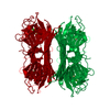
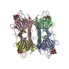
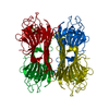
 Canavalia brasiliensis (Brazilian jackbean)
 X-RAY DIFFRACTION /
  Authors
 Citation
 Structure visualization
 Downloads & links
 Other downloads


 PDBj
PDBj


 Assembly


 Mass: 40.078 Da / Num. of mol.: 1 / Source method: obtained synthetically / Formula: Ca
 Mass: 54.938 Da / Num. of mol.: 1 / Source method: obtained synthetically / Formula: Mn
 Mass: 135.127 Da / Num. of mol.: 1 / Source method: obtained synthetically / Formula: C5H5N5
 Mass: 92.094 Da / Num. of mol.: 2 / Source method: obtained synthetically / Formula: C3H8O3
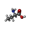
 Type: D-peptide linking / Mass: 103.120 Da / Num. of mol.: 1 / Source method: obtained synthetically / Formula: C4H9NO2
 Mass: 62.068 Da / Num. of mol.: 2 / Source method: obtained synthetically / Formula: C2H6O2
 Sample preparation
 / Beamline: D03B-MX1 / Wavelength: 1.43 Å
 Processing