 Movie
Movie Controller

[English] 日本語
 Yorodumi
- PDB-1jw6: Crystal Structure of the Complex of Concanavalin A and Hexapeptide -
+ Open data

- Basic information
| Entry | Database: PDB / ID: 1jw6 | ||||||
|---|---|---|---|---|---|---|---|
| Title | Crystal Structure of the Complex of Concanavalin A and Hexapeptide | ||||||
 Components Components |
| ||||||
 Keywords Keywords | SUGAR BINDING PROTEIN / Complex with Hexapeptide | ||||||
| Function / homology |  Function and homology information Function and homology informationregulation of defense response to virus / D-mannose binding / defense response / metal ion binding Similarity search - Function | ||||||
| Biological species |   Canavalia ensiformis (jack bean) Canavalia ensiformis (jack bean)synthetic construct (others) | ||||||
| Method |  X-RAY DIFFRACTION / MOLECULAR REPLACEMENT / Resolution: 1.93 Å X-RAY DIFFRACTION / MOLECULAR REPLACEMENT / Resolution: 1.93 Å | ||||||
 Authors Authors | Zhang, Z. / Qian, M. / Huang, Q. / Jia, Y. / Tang, Y. | ||||||
 Citation Citation | |||||||
| History |
|
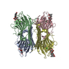
- Structure visualization
| Structure viewer | Molecule: MolmilJmol/JSmol |
|---|
- Downloads & links
-Download
| PDBx/mmCIF format | 1jw6.cif.gz | 64.1 KB | Display | PDBx/mmCIF format |
|---|---|---|---|---|
| PDB format | pdb1jw6.ent.gz | 45.3 KB | Display | PDB format |
| PDBx/mmJSON format | 1jw6.json.gz | Tree view | PDBx/mmJSON format | |
| Others |  Other downloads Other downloads |
-Validation report
| Arichive directory | https://data.pdbj.org/pub/pdb/validation_reports/jw/1jw6ftp://data.pdbj.org/pub/pdb/validation_reports/jw/1jw6 | HTTPS FTP |
|---|
-Related structure data
| Related structure data | 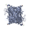 1hqwS S: Starting model for refinement |
|---|---|
| Similar structure data |


-Links
 PDBj
PDBj


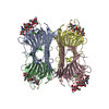
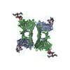
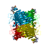
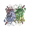
- Assembly
| Deposited unit | 
| ||||||||
|---|---|---|---|---|---|---|---|---|---|
| 1 | 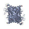
| ||||||||
| 2 | 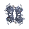
| ||||||||
| Unit cell |
| ||||||||
| Components on special symmetry positions |
|
-Components
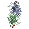
-Protein / Protein/peptide , 2 types, 2 molecules AB
| #1: Protein | Mass: 25622.385 Da / Num. of mol.: 1 / Source method: isolated from a natural source / Source: (natural) Canavalia ensiformis (jack bean) / References: UniProt: P02866 |
|---|---|
| #2: Protein/peptide | Mass: 922.057 Da / Num. of mol.: 1 / Source method: obtained synthetically / Details: This sequence coming from polypeptide libery. / Source: (synth.) synthetic construct (others) |
-Non-polymers , 5 types, 99 molecules 


| #3: Chemical | ChemComp-MN /  Mass: 54.938 Da / Num. of mol.: 1 / Source method: obtained synthetically / Formula: Mn Mass: 54.938 Da / Num. of mol.: 1 / Source method: obtained synthetically / Formula: Mn |
|---|---|
| #4: Chemical | ChemComp-CA /  Mass: 40.078 Da / Num. of mol.: 1 / Source method: obtained synthetically / Formula: Ca Mass: 40.078 Da / Num. of mol.: 1 / Source method: obtained synthetically / Formula: Ca |
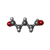
| #5: Chemical | ChemComp-PTD /  Mass: 100.116 Da / Num. of mol.: 1 / Source method: obtained synthetically / Formula: C5H8O2 Mass: 100.116 Da / Num. of mol.: 1 / Source method: obtained synthetically / Formula: C5H8O2 |
| #6: Chemical | ChemComp-IPA /  Mass: 60.095 Da / Num. of mol.: 1 / Source method: obtained synthetically / Formula: C3H8O Mass: 60.095 Da / Num. of mol.: 1 / Source method: obtained synthetically / Formula: C3H8O |
| #7: Water | ChemComp-HOH / Mass: 18.015 Da / Num. of mol.: 95 / Source method: isolated from a natural source / Formula: H2O |
-Experimental details
-Experiment
| Experiment | Method: X-RAY DIFFRACTION / Number of used crystals: 1 |
|---|
- Sample preparation
| Crystal | Density Matthews: 2.3 Å3/Da / Density % sol: 46.5 % | ||||||||||||||||||||||||||||||||||||
|---|---|---|---|---|---|---|---|---|---|---|---|---|---|---|---|---|---|---|---|---|---|---|---|---|---|---|---|---|---|---|---|---|---|---|---|---|---|
| Crystal grow | Temperature: 300 K / Method: vapor diffusion, hanging drop / pH: 5.45 Details: Sodium phosphate , pH 5.45, VAPOR DIFFUSION, HANGING DROP, temperature 300.0K | ||||||||||||||||||||||||||||||||||||
| Crystal grow | *PLUS Temperature: 25 ℃ | ||||||||||||||||||||||||||||||||||||
| Components of the solutions | *PLUS
|
-Data collection
| Diffraction | Mean temperature: 298 K |
|---|---|
| Diffraction source | Source: ROTATING ANODE / Type: RIGAKU RU300 / Wavelength: 1.5418 Å |
| Detector | Type: RIGAKU RAXIS IIC / Detector: IMAGE PLATE / Date: Oct 22, 1998 |
| Radiation | Monochromator: GRAPHITE / Protocol: SINGLE WAVELENGTH / Monochromatic (M) / Laue (L): M / Scattering type: x-ray |
| Radiation wavelength | Wavelength: 1.5418 Å / Relative weight: 1 |
| Reflection | Resolution: 1.93→25 Å / Num. all: 17973 / Num. obs: 15289 / % possible obs: 87.1 % / Observed criterion σ(F): 1 / Observed criterion σ(I): 1 / Redundancy: 4.5 % / Biso Wilson estimate: 15 Å2 / Rmerge(I) obs: 0.0527 / Net I/σ(I): 14.5 |
| Reflection shell | Resolution: 1.93→2.02 Å / Redundancy: 3.2 % / Rmerge(I) obs: 0.127 / Mean I/σ(I) obs: 4 / Num. unique all: 1961 / % possible all: 80.7 |
| Reflection | *PLUS Num. measured all: 71922 |
| Reflection shell | *PLUS Lowest resolution: 2.51 Å / % possible obs: 80.7 % |
- Processing
| Software |
| ||||||||||||||||||||||||||||||||||||||||||
|---|---|---|---|---|---|---|---|---|---|---|---|---|---|---|---|---|---|---|---|---|---|---|---|---|---|---|---|---|---|---|---|---|---|---|---|---|---|---|---|---|---|---|---|
| Refinement | Method to determine structure: MOLECULAR REPLACEMENT Starting model: PDB ENTRY 1HQW Resolution: 1.93→62.01 Å / Isotropic thermal model: Isotropic / Cross valid method: THROUGHOUT / σ(F): 1 / Stereochemistry target values: Engh & Huber
| ||||||||||||||||||||||||||||||||||||||||||
| Refinement step | Cycle: LAST / Resolution: 1.93→62.01 Å
| ||||||||||||||||||||||||||||||||||||||||||
| Refine LS restraints |
| ||||||||||||||||||||||||||||||||||||||||||
| LS refinement shell |
| ||||||||||||||||||||||||||||||||||||||||||
| Software | *PLUS Name: X-PLOR / Version: 3.1 / Classification: refinement | ||||||||||||||||||||||||||||||||||||||||||
| Refinement | *PLUS σ(F): 1 / % reflection Rfree: 9.9 % / Rfactor all: 0.21 / Rfactor obs: 0.19 / Rfactor Rfree: 0.25 / Rfactor Rwork: 0.19 | ||||||||||||||||||||||||||||||||||||||||||
| Solvent computation | *PLUS | ||||||||||||||||||||||||||||||||||||||||||
| Displacement parameters | *PLUS | ||||||||||||||||||||||||||||||||||||||||||
| Refine LS restraints | *PLUS
| ||||||||||||||||||||||||||||||||||||||||||
| LS refinement shell | *PLUS Rfactor Rfree: 0.235 / Rfactor Rwork: 0.153 |
